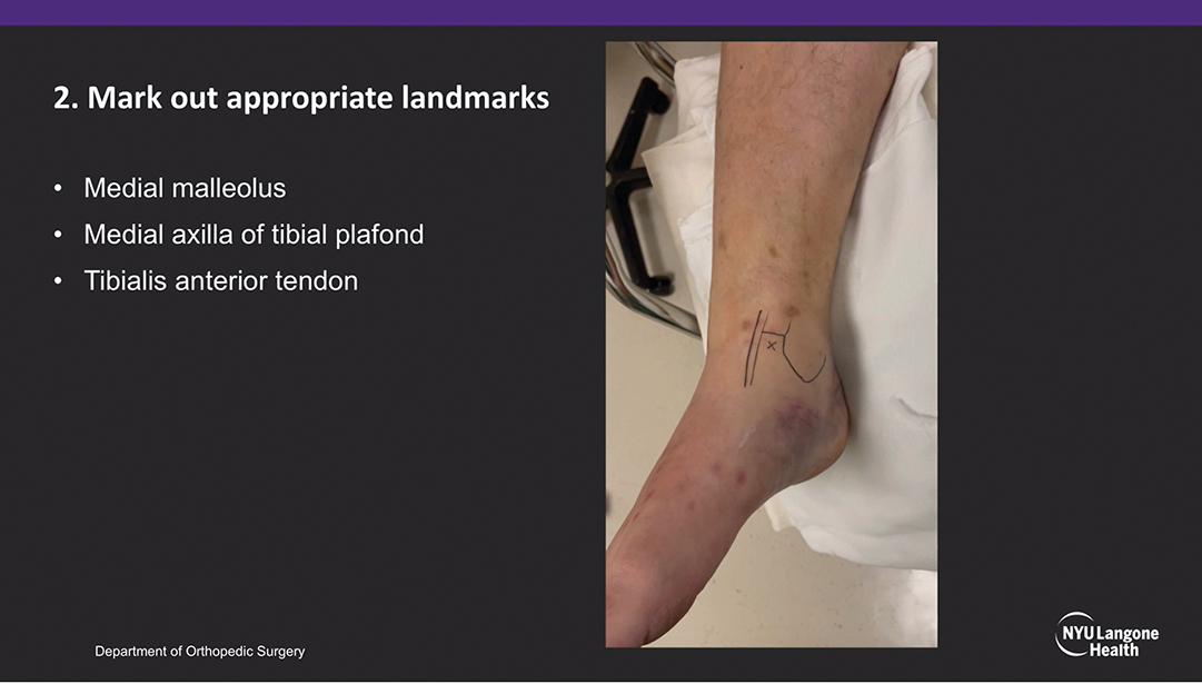
Fig. 1 Andrew Sheng Bi, MD, and colleagues mark anatomic landmarks on a patient’s ankle prior to application of an anteromedial ankle hematoma block.
Published 2/26/2025
|
Neil Jain, MD; Michael DeRogatis, MD, MS
Editor’s note: The following article is a review of a video available via the AAOS Orthopaedic Video Theater (OVT). AAOS Now routinely reviews OVT Plus videos, which are vetted by topic experts and offer CME. For more information, visit aaos.org/OVT.
Ankle fractures are common presentations, often resulting from twisting or rotational injuries. In acute settings of talar subluxation, closed reduction is often attempted to reduce deformity and relieve pressure on the skin and surrounding soft tissues. Several options are available to minimize patient discomfort during reduction, including hematoma blocks, nerve blocks, and conscious sedation with intravenous narcotic medication. Among those choices, hematoma blocks are simple, localized procedures that minimize the risks associated with stronger analgesia. Complications are rare but can include infection, cartilage damage, bleeding, or lidocaine toxicity. An understanding of anatomic landmarks and utilization of proper technique are paramount to effective relief of symptoms.
The AAOS OVT video titled “Anteromedial Ankle Hematoma Block,” from Andrew Sheng Bi, MD, and colleagues, demonstrates the steps involved in applying an anteromedial ankle hematoma block. It discusses the technique while highlighting the relevant anatomic landmarks.
The video authors start with an overview of the indications before reviewing the anteromedial space of the tibiotalar joint that is used for injection. It is noted that this space is between the medial malleolus and tibialis anterior tendon. Several risks are discussed, but the need for aspiration prior to injection is emphasized to avoid toxicity associated with direct injection into a vascular structure. Supplies needed are 10 cc of 1 to 2 percent lidocaine without epinephrine, ethyl chloride spray, an 18-gauge needle, two 10 cc syringes, ChloraPrep or alcohol swabs, 4×4 gauze, and a bandage.
To begin block application, the patient’s ankle is allowed to sit naturally in plantar flexion to facilitate a larger joint entry point. Anatomic landmarks such as the medial malleolus, medial axilla of the tibial plafond, and tibialis anterior tendon are marked (Fig. 1). The soft spot for entry into the anteromedial space is palpated between these structures. Topical ethyl chloride spray is applied at this site, and sterile preparation is performed. An 18-gauge needle enters at a slight lateral trajectory. As the needle advances, losses of resistance are expected at the levels of the skin and joint capsule. Hard stops are indicative that an adjustment in trajectory is needed.
After hematoma aspiration, the syringe is detached, and the needle is left in place. A 10 cc syringe with lidocaine is then attached to the needle. To ensure that direct injection into a vascular structure does not occur, the site is re-aspirated, and dark hematoma is expected. The lidocaine is injected, and the needle is removed. After this final step, the extremity is ready for reduction and immobilization.
Overall, this video provides a clear and detailed demonstration of an anteromedial ankle hematoma block that is of high educational value to orthopaedic surgeons and residents taking emergency department call.
Neil Jain, MD, is a postdoctoral orthopaedic surgery research fellow at St. Luke’s University Health Network in Bethlehem, Pennsylvania.
Michael DeRogatis, MD, MS, is an orthopaedic surgery resident at St. Luke’s University Health Network in Bethlehem, Pennsylvania. He is also a former member of the AAOS Now Editorial Board.
Video details
Title: Anteromedial Ankle Hematoma Block
Authors: Andrew Sheng Bi, MD; Joseph Xavier Robin, MD; Dylan Lowe, MD
Published: July 20, 2023
Time: 4:57
Tags: Featured, Trauma, Foot and Ankle, Anteromedial, Arthritis, Fracture, Pain Control
Visit aaos.org/OVT to view this award-winning title and more than 1,600 other videos from across orthopaedic topics, institutions, practice management, and industry.