Being able to identify where a tumor ends and normal tissue begins is paramount to the work of tumor surgeons. How can surgeons “see” better when doing these types of surgery? Intraoperative computer navigation and cutting guides help on a macroscopic level, but microscopically, the tools are lacking. Indocyanine green (ICG) is a water-soluble fluorescent dye that was developed during World War II for color imaging. In the 1950s, it was used to measure cardiac and renal function; in the 1960s, it was used to diagnose subretinal eye diseases. Today, its use in medicine has spread to numerous realms, including localizing tumors, assessing bowel anastomosis, lymphatic mapping, and performing muscle-flap reconstruction. In addition, several other fluorescent probes are being utilized. The application of fluorescence in orthopaedics and specifically sarcoma surgery is evolving.
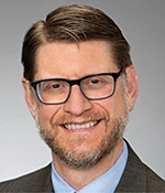
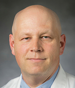
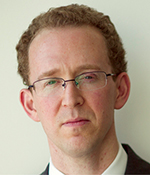
For AAOS Now, three leading experts in the field gathered to discuss this topic: Kurt Weiss, MD, FAAOS, associate professor of orthopaedic surgery at the University of Pittsburgh; Brian Brigman, MD, PhD, FAAOS, professor of orthopaedic surgery at Duke University; and Eric Henderson, MD, FAAOS, associate professor of orthopaedics at the Geisel School of Medicine at Dartmouth. Leading the discussion was AAOS Now Editorial Board member Odion Binitie, MD, FAAOS.
Dr. Weiss: My use of imaging technology has taken a circuitous route. It started out as a way to try to visualize pulmonary metastasis in my mouse model of osteosarcoma. I could not get the CT scanner to work and did not have the money for an intravascular ultrasound machine or a mouse positron emission tomography scanner. My lab resident, Mitch Fourman, MD, suggested I try ICG fluorescence imaging, so I did, and it worked. So, I thought, great, I can use this for my basic and translational science research for osteosarcoma metastasis. And that was that. However, another student proposed using it to look at intraoperative evaluation of sarcoma margins, and his study was very interesting and very successful. In my lab, we have been looking at ICG fluorescence imaging intraoperatively, to help us determine if there’s concern for a positive margin in soft-tissue sarcoma surgery. When we started the trial, I thought we were going to compare the surgeon with the machine and see which one’s better, and it turns out that was probably stupid because the machine and the surgeon are good at different things. The detector can see things that you can’t see, but you know things because of your experience as a surgeon that the machine doesn’t know. For example, you know where in the surgical field you need to be concerned with. As we go forward and hopefully build toward a randomized, multicenter study, it’s going to be combining the best of what the surgeon can do and the best of what the machine can do.
Dr. Brigman, can you tell us about your work in using a tissue-activated fluorescent probe?
Dr. Brigman: It’s important to look at why there is a problem. We do not do a good job of assessing margins. Now, what do I mean by that? If you take patients with sarcomas and treat them with surgery alone, margin status determined by pathologists does correlate with local recurrence, but it doesn’t do it very well. The specificity is about 85 percent. The sensitivity is only about 20 percent. We don’t have a good way to determine who’s going to have a local recurrence, and then we give them all radiation, and we know about the problems associated with radiation. If we could find a better way to determine if there’s sarcoma still in the patient at the time we do the surgery, we might be able to do surgeries that take less normal tissue and might be able to avoid radiation in the 60 percent of patients who get it and don’t need it.
The technology I’m using is something that was invented by David Kirsch, MD, PhD, utilizing a mouse model of soft-tissue sarcoma. It is a fluorescent probe that is activated by sarcoma and other cancers. [This technology] is a protease-activated probe. Sarcomas and other tumors have a substantial increase in protease activity compared to normal tissue. The probe is not visible and is quenched in its normal state. But in the presence of proteases, the probe breaks apart and becomes unquenched and fluorescently visible.
With this work, we could predict local recurrence far better than our pathologists could by looking at our margins. We went on to do a study in naturally occurring sarcomas in dogs. This [study] led to our first in-human phase I trial in patients with sarcoma: 15 patients, 12 of whom had sarcomas and 3 of whom had breast cancer. We were able to prove that it was safe and that we could image these tumors in human patients.
Once we were able to show safety of the technology in humans, the company was interested in moving on with a trial in breast cancer, and sarcoma became an afterthought for them. They have recently completed that trial and, in fact, got FDA approval in breast cancer. That’s where the study is so far. I’m working on putting together a multi-institutional study of soft-tissue sarcomas in humans, but it is not yet off the ground.
Dr. Henderson, tell us about your work taking fluorescence into other aspects of orthopaedics.
Dr. Henderson: Fluorescence-guided surgery is a very exciting area, utilizing probes with various mechanisms. The work that I’ve undertaken so far started with sarcoma and was aimed at improving surgical accuracy and real-time margin assessment. We’ve done work trying to synergize molecular-targeted probes using MRI- and CT-based surgical navigation and have shown that when you use all three in concert, at least in a lab-based phantom model, it improves the accuracy of navigation. Our team also has an R01 grant working to translate a nerve-specific probe.
The application that I’ve pursued most recently, however, is not cancer-related. We had a run of necrotizing fasciitis patients a few years ago, and I was frustrated by our inability to diagnose this disease correctly and immediately. It led me to research key features of necrotizing fasciitis on a biochemical level in the literature. I realized that the profound thrombosis that occurs with necrotizing soft-tissue infections could potentially be leveraged against the disease by using a perfusion probe. I started a pilot study where we enrolled 14 patients who were being evaluated for necrotizing soft-tissue infections in our emergency department. Ultimately, what we are talking about is machine recognition of tissues, to not only improve our ability to perform surgery but to see how it can also improve the ability of machines to do this. There is a lot of government funding going into research in these technologies, because they have the promise to improve surgical safety, efficacy, and access. Ultimately, I think they will be used to produce machines that will probably perform surgery better than we do.
Do you think machines are just going to take over doing surgery completely?
Dr. Henderson: I think we’re a few decades away from that, and frankly, in some of the conversations I’ve had with industry around the possibility of creating machines for surgical decision-making, due to the potential legal exposure they would take on, it is unlikely that is a direction they are willing to go.
It sounds like some of this technology is either already available or close to being available, but it’s still not, at least in sarcoma, being used at all in a real-life setting. Is that correct?
Dr. Henderson: I would agree with that. However, the leading surgical fluorescence-guidance researchers in the country and worldwide are focused on delineating contrast thresholds that indicate either, yes: tumor or no: tumor, or whatever disease, anatomy, or process you are attempting to identify. Giving the same dose of ICG to two different patients will not result in the same contrast values, even in two patients with the same diagnosis, so it’s not as simple as just injecting someone with ICG and then having an automatic and correct result given back to you. For every application, you must do the due diligence, both in the lab and in human trials, to find the thresholds and/or contrast values to accurately define what is what; it’s really more about contrast than absolute values.
Dr. Weiss, is that sort of the work that you’re doing with your ongoing trial?
Dr. Weiss: We are trying. Eric’s a million percent right. We are going to find layers and layers of complexity that have to do with the size of the tumor, the time between the infusion of the ICG and the removal of the tumor, the histologic subtype, and was radiation given? Right now, I would say we’re just crawling at best and trying to figure out how to use this intelligently. But I think between Brian, Eric, and I, and other investigators, we’re satisfied enough that we believe that it’s worth accruing more patients and trying to figure out how to use this technology in an intelligent fashion that is going to help our patients.
This is all very extremely exciting work. What’s the future?
Dr. Weiss: For me, the question is where does it really shine? Where does it really help us? Is it as good with patients who have gotten preoperative radiotherapy? Is it as good in patients who have had neoadjuvant chemotherapy? We don’t know any of the answers to those questions yet. We know the tumors appear to take it up, and there seems to be a correlation, at least in our hands, between the machine’s ability to predict local recurrence and what actually happens. So far, in a small number of patients, 54 to be exact, the detector is better at predicting local recurrence than you and I. How do we use this in an intelligent way? Where is it going to be the best, and what are the tricks of the trade that we need to apply to sort of maximize it? I feel there’s a certain nihilism in our field that local recurrences are going to happen, they are a part of doing business, and I might agree with that, but I don’t accept the hypothesis that we can’t do better. That’s where I hope things go in the next 5 to 10 years.
Dr. Brigman, what’s next for you? You mentioned that you have an upcoming trial utilizing the Lumicell probe specifically in sarcoma.
Dr. Brigman: I have submitted the trial for funding through the R01 mechanism of the National Institutes of Health for a feasibility study in soft-tissue sarcomas. We will be looking at the wide variety of soft-tissue sarcoma subtypes, looking to see if they are all imageable. We will assess margins with the system, trying to determine the exact sensitivity and specificity of the system for detecting tumor—that would be the goal of the study, which would hopefully inform a true clinical trial that would happen subsequent to that.
Dr. Henderson, what are the next steps in your work in necrotizing fasciitis?
Dr. Henderson: Our forthcoming R01 grant is aimed at using ICG as a diagnostic agent. After that, the next step is to use it as a surgical guidance tool for necrotizing fasciitis, to in real time plan out your incisions to remove the entire area affected, much like obtaining negative margins for sarcoma. From a more global standpoint, we need to have a more effective way to get these probes to market. The FDA oversees the approval of the probes and the devices that detect the probes. There is tremendous uncertainty about how these drugs and devices should be regulated because the drugs are not being applied therapeutically and it’s unclear whether the probe or the device is the key element in making a diagnosis or guiding the surgery. The ultimate regulatory pathway will likely be similar to radiological contrast agents (e.g., gadolinium), but because these technologies are intended to guide surgery, it is a different beast. At present, there is not a streamlined pipeline to get these to market, and there is substantial heterogeneity in the design of trials and the metrics to evaluate performance.
Thank you all for the work you are doing and for spending the time discussing the burgeoning and exciting area in our field.
Odion Binitie, MD, FAAOS, is an associate professor of orthopaedics and oncology at Moffitt Cancer Center and the University of South Florida College of Medicine in Tampa. He is on the Editorial Board of AAOS Now.


