Editor’s note: The following article is a review of a video available via the AAOS Orthopaedic Video Theater (OVT). AAOS Now routinely reviews OVT Plus videos, which are vetted by topic experts and offer CME. For more information, visit aaos.org/OVT.
Humeral diaphysis fractures are common injuries, most often resulting from direct trauma or low-energy falls. A careful physical examination on presentation is necessary due to a risk of neurovascular deficits, usually involving the radial nerve. When inspected, a limb may present with acute shortening and a varus deformity due to underlying deforming forces.
Management of simple fracture patterns is typically nonoperative, with placement of a coaptation splint for 7 to 10 days. After resolution of acute-phase symptoms, patients can be transitioned to a functional brace or undergo operative management if necessary. Patients who should be considered for nonoperative treatment include those with long oblique or spiral fracture patterns with acceptable alignment after reduction, closed injuries or low-velocity gunshot wounds, and no vascular or brachial plexus injury, as well as patients who are not morbidly obese or have significant comorbidities precluding operative management.
There is no consensus on what constitutes acceptable alignment, but the following criteria have been proposed: 20 degrees of anterior or posterior angulation, 30 degrees of varus or valgus angulation, 15 degrees of malrotation, and 3 cm of shortening. Transverse and short oblique fractures have minimal boney contact and therefore are at higher risk of non-union.
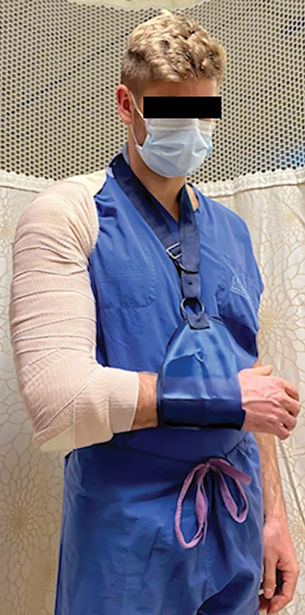
Although most humeral shaft fractures can initially be treated non-operatively, some patients may eventually require surgical fixation. To help mitigate surgery in select patients, proper reduction and comfortable coaptation splinting can be performed in the emergency department setting. The AAOS OVT video titled “Coaptation Splinting: Modified Method” demonstrates the steps involved in the placement of a coaptation splint. It discusses the indications and technique for immobilization while highlighting management after placement.
The authors start with a careful review of the indications and workup to be completed prior to immobilization. They emphasize the need for careful physical examination and assessment of imaging due to the risk of neurovascular injury associated with humeral shaft fractures. They note that a coaptation splint is applied in the acute phase to allow symptoms such as swelling to subside before transition to a brace or surgical intervention.
To begin splint application, measure plaster from the axilla, around the elbow, and back up to the level of the neck on the uninjured arm. Then cut a stockinette to 1.5 times the previously measured plaster. At one end, approximately one-third of the stockinette is cut in half.
Layer 10 strips of plaster and then sandwich them between six layers of cotton padding on each side. Remove excess material, and pull the plaster construct into the stockinette.
To apply the splint, place the construct high onto the axilla and tie the two strands of the previously cut stockinette around the contralateral side of the neck. Then drape the other side of the plaster construct over the arm. Retie the stockinette strands over the ipsilateral shoulder and secure the splint with a compressive wrap. It is recommended to start above the elbow, work distally, and then return proximally. Apply a valgus mold to counteract the deforming forces of humeral shaft injuries typically causing varus extension. Conduct a post-splint neurovascular exam, and finalize the splint wrap after removal of the previously tied stockinette knot. Then fashion the patient in a cuff and collar. The authors explain that post-splinting protocol involves Sarmiento bracing after swelling resolution, early gradual range of motion, and weekly follow-up.
Overall, this OVT video is a clear and detailed demonstration of coaptation splint placement that is of high educational value to orthopaedic residents and junior orthopaedic surgeons.
Michael DeRogatis, MD, MS, is an orthopaedic surgery resident at St. Luke’s University Health Network in Bethlehem, Pennsylvania, and serves as a resident member of the AAOS Now Editorial Board.
Neil Jain, MD, is a postdoctoral orthopaedic surgery research fellow at St. Luke’s University Health Network in Bethlehem, Pennsylvania.
Video details
Title: Coaptation Splinting: Modified Method
Authors: Zachary Mills, MD; Nathan Benner, MD; Eli W. Bunzel, MD; Arien Lee Cherones; Robert Paul Dunbar Jr, MD, FAAOS
Published: Feb. 27, 2024
Time: 10:47
Tags: Trauma, Humeral Shaft Fracture, Casting and Fracture Bracing, CME
Visit aaos.org/OVT to view this award-winning title and more than 1,600 other videos from across orthopaedic topics, institutions, practice management, and industry.