Radiofrequency echographic multi-spectrometry (REMS) is an ultrasound technology that assesses bone mineral density (BMD) and bone quality of the lumbar spine and hips. The predecessor of REMS technology, quantitative ultrasound (QUS), was found to have clinical relevance as a predictor of fracture risk. However, due to significant variability in the sensitivity and specificity of QUS in BMD, it could not be used as a clinically valid method to diagnose and treat osteoporosis based on the World Health Organization (WHO) guidelines. REMS technology has overcome the limitations of QUS. Advancements in ultrasound technology have resulted in the development of innovative hardware and software that allow for assessment of BMD and bone quality at the regions of interest in the lumbar spine and hip. The results are rapidly reported in real time and immediately available to both the examiner and patient.
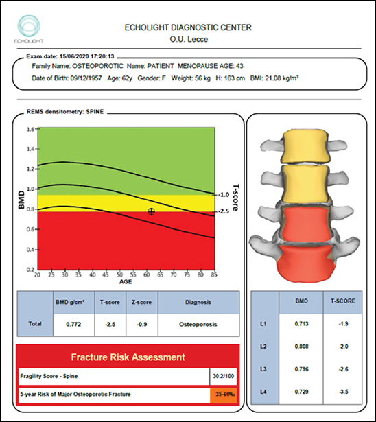
A transducer is applied to the region of interest (ROI), then ultrasound waves are emitted and interact with the targeted bone tissue in the ROI. The backscatter radiofrequency signal, or “echo,” is captured for analysis, yielding an osteoporosis score (OS). The OS value is then statistically analyzed by Pearson correlation coefficient and compared to a proprietary reference database classified as normal density, osteopenic (low bone density), or osteoporotic. The OS is used to calculate BMD with corresponding T-score and Z-score (Fig. 1). This approach has been validated across multiple clinical studies.

REMS is comparable to dual-energy X-ray absorptiometry (DXA) in determining BMD and generating T-scores and Z-scores that are clinically significant and valid in diagnosing osteoporosis. Several reference parameters have been reported that document high accuracy, reproducibility, and precision of REMS technology. Artifacts such as osteophytes, vertebral fractures, and metal structures are automatically excluded by the algorithm, thereby avoiding potential for error in the estimation of BMD.
Pisani et al demonstrated that REMS accuracy and precision were comparable to DXA, signifying its effective clinical use for the diagnosis of osteoporosis according to WHO criteria. REMS can be used to monitor BMD and provide information that is accurate, precise, and reproducible.
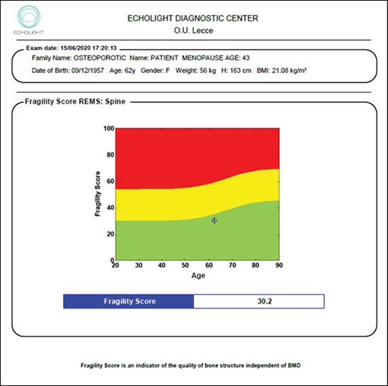
The most compelling aspect of REMS technology is the ability to assess bone quality independent of BMD to provide a predictive tool for fracture risk, referred to as the Fragility Score (FS) (Fig. 2). The FS has shown the ability to discriminate between patients who experienced an osteoporotic fracture and those who did not, with sensitivity and specificity of 76 percent and 68 percent, respectively, compared with sensitivity of 73 percent and specificity of 68 percent in DXA BMD. In a 5-year longitudinal study, Pisani et al found that FS is a better predictor of fracture risk than BMD obtained from either DXA or REMS, confirming the importance of bone quality in fracture risk determination.
REMS can be used to assess bone health in clinical situations when ionizing radiation may not be appropriate, such as pregnancy. REMS has been shown to be more accurate than DXA for monitoring bone health in individuals with type 2 diabetes mellitus (T2DM). A 2020 study by Caffarelli et al found that DXA showed higher bone density measurements in women with T2DM than in nondiabetic patients, whereas REMS showed lower measurements. REMS classified a higher percentage of T2DM women as osteoporotic. REMS showed significant differences in BMD values between those with and without major fragility fractures. REMS can be used in the evaluation of patients with skeletal deformities (e.g., osteogenesis imperfecta) and/or in the presence of orthopaedic instrumentation where the hardware will adversely affect the results of a DXA study. In such cases, REMS technology is a critical tool for monitoring bone health status, documenting disease progression, and monitoring treatment effect. Furthermore, insurance approvals for the appropriate osteoporosis therapy are often based on BMD measurements that cannot be reliably received in many patients, such as those with significant bone deformities or presence of instrumentation.
REMS is a groundbreaking technology that can raise osteoporosis awareness and increase osteoporosis screening due to high-quality, reliable, and readily accessible technology. The absence of ionizing radiation and the portable nature of REMS technology allow for osteoporosis diagnosis at the point of care more broadly and at an earlier stage. The tool also enables accurate monitoring after diagnosis. These features—as well as others, such as ease of use, accurate and reproducible results, and the possibility of bedside scanning—when combined with economic advantages of a less costly system and the cost savings from earlier detection and treatment of osteoporosis, can help expand access to high-quality musculoskeletal healthcare and reduce the risk of fracture for people everywhere.
Kimberly Zambito, MD, FAAOS, FAOA, is an orthopaedic hand surgeon at St. Luke’s University Health Network in Bethlehem, Pennsylvania.
Andrew Bush, MD, FAAOS, FACS, is an orthopaedic surgeon at Central Carolina Orthopaedics Associates in Sanford, North Carolina.
Michael DeRogatis, MD, MS, is an orthopaedic surgery resident at St. Luke’s University Health Network in Bethlehem, Pennsylvania, and serves as a resident member of the AAOS Now Editorial Board.
References
- Hans D, Baim S: Quantitative ultrasound (QUS) in the management of osteoporosis and assessment of fracture risk. J Clin Densitom 2017;20(3):322-33.
- Conversano F, Franchini R, Greco A, et al: A novel ultrasound methodology for estimating spine mineral density. Ultrasound Med Biol 2015;41(1):281-300.
- Casciaro S, Peccarisi M, Pisani P, et al: An advanced quantitative echosound methodology for femoral neck densitometry. Ultrasound Med Biol 2016;42(6):1337-56.
- Pisani P, Conversano F, Muratore M, et al: Fragility score: a REMS-based indicator for the prediction of incident fragility fractures at 5 years. Aging Clin Exp Res 2023;35(4):763-73.
- Cortet B, Dennison E, Diez-Perez A, et al: Radiofrequency echographic multi spectrometry (REMS) for the diagnosis of osteoporosis in a European multicenter clinical context. Bone 2021;143:115786.
- Fuggle N, Reginster JY, Al-Daghri N, et al: Radiofrequency echographic multi spectrometry (REMS) in the diagnosis and management of osteoporosis: state of the art. Aging Clin Exp Res 2024;36(1):135.
