The Surgical Approach to The Distal Medial Femur
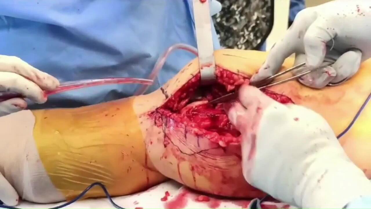
Introduction: The surgical approach to the distal medial femur is ideal for open reduction and internal fixation of articular distal femoral fractures with a medially based apex. Placing a plate in an anti-glide fashion at the fracture apex allows for the most stable construct in this load-bearing region. In these cases, an anatomic reduction of the articular surface is achieved not only at the joint level but also at the apex of the fracture. We present a case of simple distal medial condyle sagittal fracture through the intercondylar notch (AO/OTA 33 B2.1) in a 60-year-old female, that was surgically managed with open reduction and internal fixation, using an anatomic medial distal femoral locking plate. We aim to highlight our surgical approach to the distal medial femur and emphasize our technique of ORIF for the distal medial femoral condyle.Surgical Technique: The patient is positioned supine on an orthopedic radiolucent table, and towels are placed under the thigh to keep the ipsilateral hip and knee slightly flexed. The skin incision is marked as a director medial approach along the aspect of the distal femur, extending from the level of the joint to approximately 15 to 20 centimeters proximally. The skin is incised, and the incision is carried down through deep fatty tissues to the medial fascia overlying the Vastus Medialis Obliquus (VMO). The fascia is exposed in a limited fashion anteriorly and posteriorly, generally with a Cobb elevator to facilitate closure. The fascia is incized and the muscle mass of the VMO is digitally dissected on its posterior border. The VMO muscle mass in its entirety is elevated and retracted anteriorly. This allows and facilitates exposure of the medial femoral cortex. The deep soft tissues and the capsule are incised at the level of the joint. A formal arthrotomy is performed to allow direct visualization of the articular surface. Note that minimal bleeding is associated with this approach, even without the use of a tourniquet, as in this case. Once the capsule is carefully incised, surgical exposure and visualization of the articular surface, debridement of interposed hematoma and soft tissues, and the articular reduction are facilitated. The periosteal elevation is limited to the proximal portion of the fracture apex as well as the distal aspect of the medial femoral cortex. Note that a full debridement of the fracture site may necessitate using a small laminar spreader. Once the fracture site has been adequately debrided, direct reduction is then performed using pointed reduction clamps, and both the intra-articular surface as well as the bony apex are evaluated to confirm the reduction. A provisional fixation is placed using K-wires to allow for the removal of the reduction clamp and to facilitate plate placement. In this case, an anatomic medial distal femoral plate was selected to allow early weight bearing and allow for locking screws distally in this osteopenic patient. The position of the plate is confirmed fluoroscopically, and K wires are used to affix the plate to the bone distally while ensuring the plate position using the C-arm. Proximally, multiple non-locking screws are placed, and the distal fixation is placed with either locking or non-locking screws. Wound closure is performed by closing deep fascial layers and then the subcutaneous layer and skin in a typical fashion. Postoperative Care: In general, the patient is immobilized for a brief period, beginning early range of motion. Weight-bearing is determined based on the patient's age, bone quality, and fracture configuration. In this presented case, immediate full weight bearing wasn't permitted.