Best Practices: Gravity Stress Views of Ankle Fractures to Determine Stability
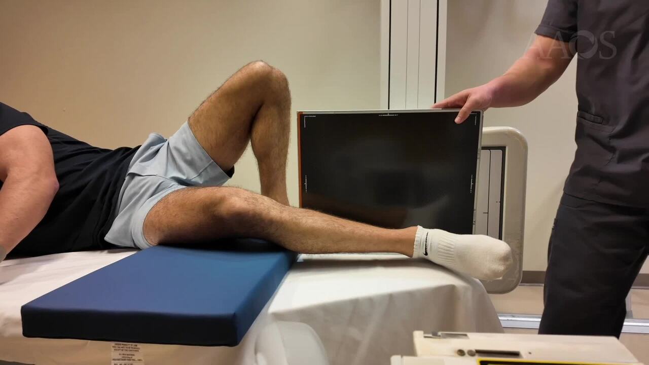
Introduction: Ankle fractures are among the most common injuries treated by orthopedic surgeons, with approximately 585,000 ankle fractures annually, 25% of which necessitate surgical intervention [1]. Gravity stress radiographs are crucial in evaluating the stability of ankle fractures, especially Weber Type B fractures that occur at the level of the distal tibiofibular syndesmosis [2,3,4]. This technique offers significant advantages, including reduced radiation exposure for medical personnel and decreased patient discomfort compared to traditional stress radiographs. Accurate assessment of fracture stability is vital for determining the need for surgical intervention and devising an effective treatment strategy. This video provides a detailed, step-by-step guide for performing a gravity stress radiograph on a patient with a suspected Weber Type B ankle fracture. The patient is positioned on the hip of the affected side, with the opposite leg used for support. A sponge pad is placed under the affected knee, allowing the injured ankle to hang approximately one inch above the bed, facilitating gravity-induced stress. The X-ray detector is placed behind the heel of the injured ankle and secured using a detector holder. The X-ray tube is aligned with a 10-degree cephalic angle from anterior to posterior to capture a mortise view. For setups with a built-in detector, the patient’s ankle hangs off the end of the table, and the X-ray tube is centered at the ankle with the detector adjusted accordingly.Conclusions: Gravity stress radiographs are particularly valuable for Weber Type B fractures due to the potential variability in stability from associated deltoid ligament ruptures or medial malleolus fractures. Mastery of proper radiographic techniques is essential for accurate stability assessment, which in turn guides effective treatment planning. This instructional video aims to improve the understanding and execution of gravity stress radiographs in clinical practice to enhance patient outcomes.ReferencesFlynn, J. M., Rodriguez-del Rio, F., & Pizá, P. A. (2000). Closed ankle fractures in the diabetic patient. Foot & ankle international, 21(4), 311–319. https://doi.org/10.1177/107110070002100407 Gill, J. B., Risko, T., Raducan, V., Grimes, J. S., & Schutt, R. C., Jr (2007). Comparison of manual and gravity stress radiographs for the evaluation of supination-external rotation fibular fractures. The Journal of bone and joint surgery. American volume, 89(5), 994–999. https://doi.org/10.2106/JBJS.F.01002 van Leeuwen, C., Haak, T., Kop, M., Weil, N., Zijta, F., & Hoogendoorn, J. (2019). The additional value of gravity stress radiographs in predicting deep deltoid ligament integrity in supination external rotation ankle fractures. European journal of trauma and emergency surgery : official publication of the European Trauma Society, 45(4), 727–735. https://doi.org/10.1007/s00068-018-0923-x van Leeuwen, C. A. T., Krijnen, P., Hoogendoorn, J. M., & Schipper, I. B. (2022). The value of radiologic diagnostics in evaluating deltoid integrity in isolated type B ankle fractures: a systematic review of the literature. Archives of orthopaedic and trauma surgery, 142(7), 1523–1530. https://doi.org/10.1007/s00402-021-03850-y