Medial Patellofemoral Ligament Reconstruction With Allograft Through Double Transverse and Converging Patellar Tunnel for Patellar Instability: From Anatomy To Surgical Treatment
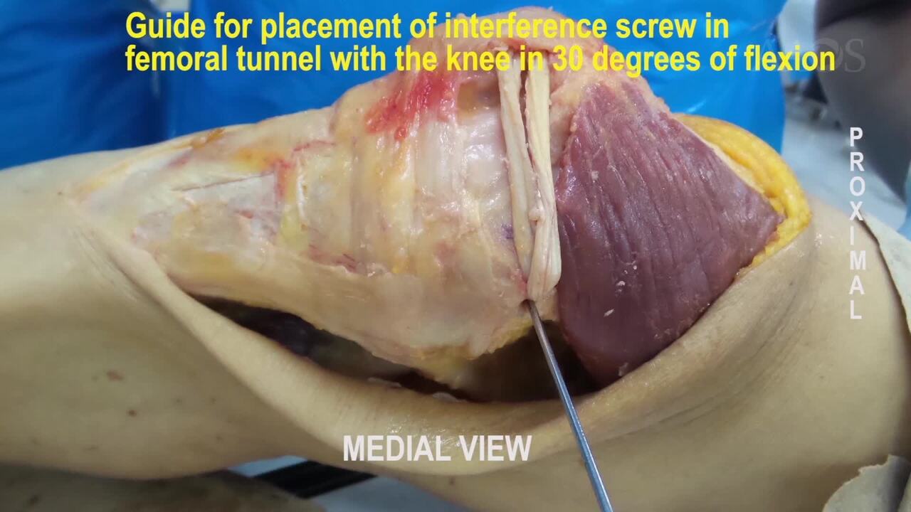
Introduction: Patellofemoral instability is one of the knee complications that can be found in up to 3% of knee injuries, especially with younger patients and female patients. After the initial dislocation, most patients will have a medial patellofemoral ligament (MPFL) injury. The etiology of the instability is multifactorial. Soft tissue and bone are the main stabilizers of physiological patellar tracking. The soft-tissue restraints contributing to patellofemoral stability consist of the vastus medialis obliquus (VMO) component of the quadriceps muscle, the medial retinaculum (the medial patellotibial/meniscal ligaments), and the medial patellofemoral ligament (MPFL). In patients with normal patellofemoral morphologic features and lower-extremity alignment, patellar instability results from deficient soft-tissue stabilizers. Several biomechanical studies showed that MPFL is It is the main restrictor of the lateral translation of the patella up to 30° of flexion. The reconstruction of the MPFL, which restores native length and stiffness of the medial soft tissue, must be the goal of a successful surgical intervention.Purpose: To show the normal anatomy of the main stabilizing soft tissues of normal patellar tracking and a video of Medial patellofemoral ligament (MPFL) reconstruction with a posterior tibial tendon allograft in cadaver lab. Additionally, we investigated the outcomes of MPFL reconstruction with this allograft through double transverse and converging patellar tunnel in our patients.Methods: A video of gross anatomy of the medial area of patellofemoral joint with its stabilizing medial soft tissues and a video of MPFL reconstruction MPFL reconstruction with a posterior tibial tendon allograft passed through double transverse and converging patellar tunnel and then the graft is passed through a tunnel in the medial femoral condyle from medial to lateral and fixed with an interference screw. The video shows the surgical procedure performed to reconstruct the MPFL in cadaver laboratory and in a patient. The rehabilitation protocol consisted of the use of crutches for 2 weeks postoperatively, quadriceps isometric exercises from the first postoperative day, allowing for progressive weight bearing and range of motion, knee flexors strengthening after two weeks, running and slowly progressive sports at 12 weeks and sports return at 24 weeks. 17 patients were reviewed, and the outcomes were assessed at a mean follow-up of 74 months (range, 42 to 136 months, standard deviation (SD) ± 2,82) with the Lysholm score and Kujala score.Results: A total of 17 patients (7 men, 10 women) with a mean age of 21.8 years (range, 15 to 32 years; standard deviation ± 4.24 years) were included in the study. All patients had recurrent lateral patellar dislocation. At the end of follow-up, the patients had patellar stability, and only one of the patients had presented an episode of subluxation at 8 months postoperatively, which did not occur again in the subsequent 5 years of follow-up. The mean Lysholm score improved from 49 preoperative (range, 33 to 71, SD deviation ± 9.19) to 81 (range, 56 to 95, SD ± 18.38) at final follow-up and the mean Kujala score improved from 53 preoperative (range, 30 to 84, SD ± 5.65) to 82 (range, 48 to 98, SD ± 12.02) at final follow-up.Conclusion: MPFL is It is the main restrictor of the lateral translation of the patella up to 30° of flexion. The reconstruction of the MPFL with allograft through double transverse and converging patellar tunnels restores patellar stability.