Thumb Radial Collateral Ligament Reconstruction with Palmaris Longus Grafting and Internal Brace Augmentation
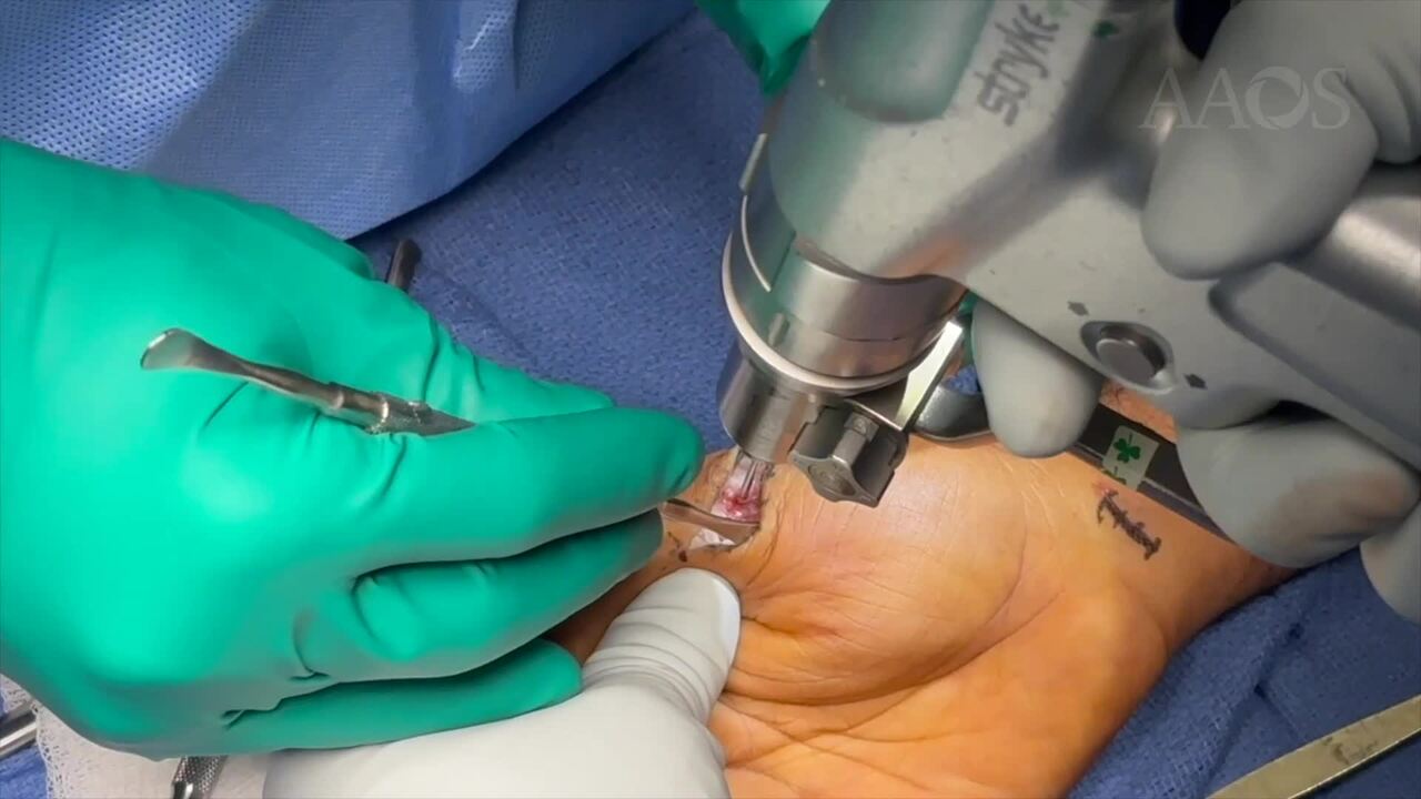
Proposal: Radial collateral ligament (RCL) injuries of the thumb are thought to be relatively uncommon injuries resulting from sudden adduction of the thumb metacarpophalangeal joint. Chronic RCL injuries contribute to significant thumb instability. Depending on tissue quality and chronicity of the injury, RCL tears may be treated with primary repair or reconstruction often using free tendon grafts, such as the ipsilateral palmaris longus tendon. The authors describe an innovative method of radial collateral ligament reconstruction with the use of a palmaris longus graft and fixation with internal brace for dynamic stabilization of the thumb metacarpophalangeal joint.Case Overview: The patient is a young adult male who sustained a right thumb injury after lifting furniture with pain localized over the radial collateral ligament. Examination demonstrates laxity of the metacarpophalangeal (MCP) joint with ulnar stress. MRI obtained confirmed a high grade partial tear of the radial collateral ligament.Method/Technique: A 2cm curvilinear incision centered over the radial collateral ligament of the MCP joint was utilized. The subcutaneous tissue was dissected to preserve the radial sensory nerve. The joint was exposed after careful elevation of the abductor aponeurosis. Upon visualization of the radial collateral ligament, it was found to be insufficient and densely scarred. Given the confirmed instability and extensive scarring, we had opted to proceed with reconstruction of the radial collateral ligament. The metacarpal neck and the base of the proximal phalanx are debrided to expose bone. The points of insertion of the free tendon graft are localized just dorsal to the rotational axis of the metacarpal head and the lateral tubercle of the proximal phalanx. Two guidewires are placed at these points of insertion to guide screw fixation. Once appropriate positioning was confirmed on fluoroscopy, attention was turned to harvesting the palmaris longus graft. The palmaris longus tendon is harvested from the ipsilateral forearm using a tendon stripper. One end of the graft was inserted into the proximal phalanx and secured with a biotenodesis screw along with a suture tape as an internal brace. After appropriate length of the tendon graft was measured and cut, the proximal end of the graft was secured with a 2-0 Fiberwire and inserted into the metacarpal neck and secured with a biotenodesis screw along with a suture tape. This provided excellent fixation and restoration of the radial collateral ligament. The patient was splinted in a thumb spica splint for 2 weeks and then transitioned to a removable splint. Immediate finger range of motion was encouraged with progressive to strengthening at 6 weeks.Results: At the patient’s 2 month postoperative visit, he was able to flex his MCPJ and no laxity was noted on examination of the radial collateral ligament. Our anatomic reconstruction using a palmaris longus tendon graft and internal brace is simple and restores RCL anatomy while providing secondary stabilization. With these techniques we were able to restore MCP flexion and stability.Summary: Thumb radial collateral ligament reconstruction using a palmaris longus tendon graft and internal brace is an effective option for the management of stabilization of thumb MCPJ instability. Studies have shown primarily excellent results using the palmaris longus tendon using the Glickel grading system with good functional results without significant donor site morbidity.