Freeing a Stiff Elbow After ORIF: An Elbow Contracture Release Technique
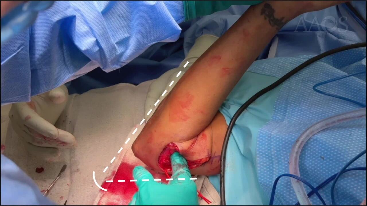
Proposal: Elbow contracture is a disabling complication frequently encountered after operative elbow injuries. Despite the satisfactory reduction and fixation, distal humerus fractures, in particular, can lead to elbow stiffness. The causes of elbow contractures include capsular thickening/contracture, heterotopic ossification, malunions, and osteophyte formation. Surgical treatment requires intimate knowledge of the anatomy, systematically targeting the contributing factors to elbow contracture. We provide strategies to circumferentially release the elbow joint in the setting of distal humerus fracture ORIF.Case Overview: This is a technique video of a compilation of open elbow contracture releases performed for patients with distal humerus fractures. All patients underwent uneventful surgical fixation followed by early motion therapy within 1-2 weeks. Despite 6 months of therapy, the elbow range of motion was limited to less than 75° arc of motion with pain at terminal flexion and extension.Method/Technique: The medial side is approached first. After the skin incision, the over-the-top approach was utilized to expose the medial column of the humerus, which serves as an important landmark. The ulnar nerve is also meticulously dissected from the scar tissue and mobilized. The brachialis and the anterior half of the flexor/pronator mass are elevated anteriorly. The capsule is released from the humerus, and approximately 1 cm width of the capsule is excised. With the anterior compartment exposed, the coronoid and the coronoid fossa can be debrided, and the brachialis can be released off the humerus proximally. The ulnar nerve is transposed anteriorly, and the posterior compartment of the elbow can be exposed by developing the plane between the medial column of the humerus and the triceps. Through this interval, the posterior capsulectomy, triceps tenolysis, and olecranon tip debridement can be performed. The posterior bundle of the medial collateral ligament (PMCL) is located on the floor of the cubital tunnel. Once the PMCL is fully released, the ulnohumeral joint should be directly visible, and significant flexion should be achieved. As long as the anterior surface of the medial epicondyle and medial border of the coronoid are left untouched, the anterior bundle of the medial collateral ligament is generally safe. The lateral incision can be shorter, and the radiocapitellar joint is directly approached using Kaplan’s approach. The anterior capsulectomy is completed laterally, and the adhesions of the brachioradialis are released off the humerus. Heterotopic ossification is also frequently encountered posterolaterally. A vast majority of the contracture release is done medially, but authors found the elbow release laterally adds 10-20° arc of motion. Implants are removed based on patient symptoms and surgeon preference. The elbow is splinted in extension to reduce the elbow joint volume and promote hemostasis. Drains are utilized in about 30% of the cases. The therapy starts on postoperative day 1-3.Results: The postoperative range was approximately 5° - 135° arc of motion. The patients commonly experience less pain with increased mobility and the resolution of ulnar nerve symptoms. Recurrent HO and elbow instability are rare. Failed contracture releases were encountered during our early experience, likely due to inadequate release.Summary: Elbow contracture is a common complication encountered after complex elbow injuries. A systematic approach to open contracture release of the elbow leads to satisfactory clinical outcomes.