One-stage Metatarsal Lengthening by Allograft Interposition for the Treatment of Complex Forefoot Deformity with Brachymetatarsia
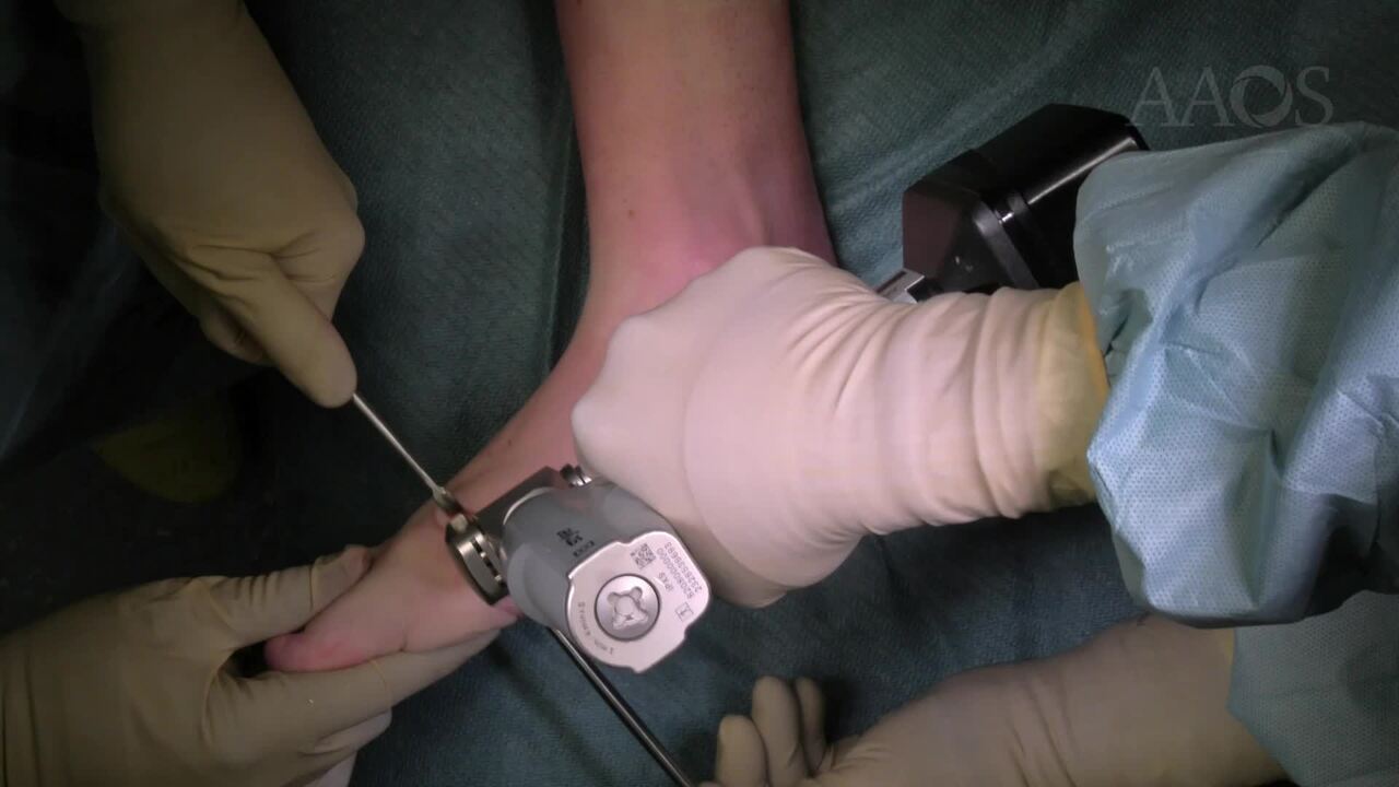
Introduction: Brachymetatarsia is a forefoot deformity consisting in one or more shortened metatarsal bones. It predominantly affects females with a high bilateral predominance. The fourth metatarsal is the mostly affected. Due to this structural anomaly, it can cause further deformities of the forefoot bones. The deformity, which may be congenital, idiopathic or secondary to surgery or trauma, may result in functional as well as cosmetic alterations. When pain occurs, conservative treatments can alleviate symptoms. However, if these methods prove ineffective, surgical intervention may be considered. Several operative options have been suggested like soft tissue release, syndactyly, one-stage or gradual lengthening of the metatarsal. The aim of this video is to describe the surgical correction of a complex forefoot deformity, comprising brachymetatarsia of the III and IV metatarsal bones and inter- and metatarsophalangeal hallux valgus by one-stage metatarsal lengthening with homologous bone graft, linear distal metatarsal (SERI) and Akin osteotomies.Materials and Methods: This study retrospectively enrolled surgical treated patients affected by brachymetatarsia. Standard weightbearing X-Rays of the foot were performed to evaluate the deformities of the forefoot. SERI and Akin osteotomies were performed, respectively, with a medial approach to the neck of the first metatarsal bone followed by a distal linear lateralizing osteotomy and a medial approach to the proximal phalanx with medial closing wedge osteotomy. The two osteotomies were then stabilized with a single K-wire inserted in a proximal to distal direction through the metatarsal incision. A dorsal longitudinal incision was made over the III intermetatarsal space, centered on the proximal metaphysis of the affected metatarsal. The extensor tendons of the affected metatarsal bones were elongated with a Z-plasty. A transverse proximal osteotomy of the metatarsals was performed and the metatarsal bone was lengthened as planned. The previously prepared bone graft was inserted. Finally, the constructs were stabilized with a K-wire. The AOFAS, MOxFQ, VAS scores and X-Rays were evaluated pre and post-operatively.Results: A total of 16 patients, 27 feet, were treated, with a mean age 27 (range 12-42), 14 females and 2 males. The mean follow-up (FU) was 3 years (range 1-5). Five patients had bilateral brachymetatarsia. Of the 27, five had multiple brachymetatarsia which involved the third and fourth metatarsals in all cases. In total we treated 37 metatarsals. All patients complained of appearance and discomfort: 11 feet had hallux valgus and 17 hammertoes. The AOFAS score improved on average from 52.2&[plusmn]7.3 (range 40-64) preoperatively to 81.6&[plusmn]4.8 (range 70-92) postoperatively. The MOxFQ score improved on average from 67.2&[plusmn]12.2 (range 50-85) preoperatively to 26.4&[plusmn]8.9 (range 18-34) postoperatively. The VAS score improved on average from 5.2&[plusmn]2.1 (range 3-7) preoperatively to 3.7&[plusmn]1.2 (range 1-5) postoperatively. On the X-Rays the mean postoperative gain in length was 13mm&[plusmn]1.1 (range 10-15.2mm) and was 12.3mm at 3 months FU. In 24 cases the metatarsal parabola was restored. Two complications were reported. One case of a bone graft mobilization that required a reintervention. One case of a partial bone graft mobilization that was treated conservatively and resulted in good consolidation over time.Discussion: Brachymetatarsia is a deformity that causes forefoot functional impairment and aesthetic problems. Surgery is not performed until the bone growth is completed and symptoms arise, and, for the time being, conservative treatments are used. When conservative methods fail, there are several surgical options that can be considered. In order to avoid neurovascular damage, lengthening of more than 15mm are treated by gradual lengthening, meanwhile when the length defect is less than 15mm, a one-stage procedure can be employed. Gradual lengthening by callus distraction with an external fixator enables elongation of both bone and soft tissue, achieving greater length increases. However, this method requires multiple surgical interventions, strong patient compliance, and nearly twice the healing time compared to one-stage procedures. One-stage lengthening typically has less impact on patients' daily routines but is limited by the extension capacity of soft tissues, restricting the attainable length gain. This procedure can be performed with autologous, homologous, or synthetic bone. Our data suggest one-stage metatarsal lengthening using metatarsal homologous bone graft is a reliable technique to correct brachymetatarsia with general satisfaction by the patients. Correction of the other toe deformities allows the right alignment of the rays and the restoration of the metatarsal parabola, thus a correct redistribution of the pressure on the metatarsal heads.Conclusions: It is not possible to identify a gold standard technique to address brachymetatarsia. The two predominant surgical techniques bear advantages and drawbacks. One-stage metatarsal lengthening using metatarsal homologous bone graft is well tolerated, with short recovery time, low morbidity, limited complications, restored forefoot anatomy and high functional scores.