Open Reduction and Internal Suture Anchor Fixation of an Isolated Lesser Tuberosity Fracture
The patient is a 27-year-old woman who sustained an isolated lesser tuberosity fracture approximately 6 weeks ago during a skiing accident. After being treated nonsurgically via a sling for 3 weeks, the patient presented to the clinic with pain and limited range of motion at the shoulder joint. Radiographs and CT scans revealed a displaced and angulated isolated lesser tuberosity fracture, with the major fracture fragment being displaced inferiorly and rotated almost 90°.
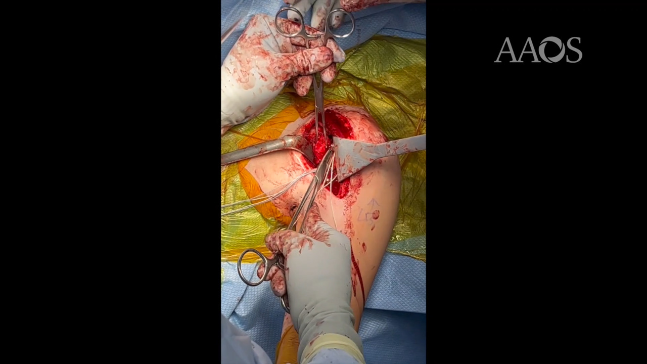
A deltopectoral approach to the humeral head was used. An incision was created following the line of the deltopectoral groove. After the cephalic vein was displaced laterally, the deltoid and pectoralis major muscles were retracted, and the humeral head was exposed. The fracture fragment had partially healed in a malunion, with the fragment being rotated 90° vertically and displaced medially. A curved osteotome was used to mobilize the fracture fragment.
The medial row was prepared using two fully threaded, cancellous 4.5-mm suture anchors, which were inserted into the medial border of the footprint of the fracture site. The suture limbs were then shuttled in a mattress fashion through the subscapularis tendon, medial to the fracture fragment. All the suture limbs were then pulled laterally to reduce the fracture. To begin lateral row fixation, the suture limbs from the medial row were passed through the islets of two cannulated and vented 4.75-mm knotless suture anchors. The lateral row suture anchors were inserted into the humeral head just lateral to the fracture site.
After sturdy fixation was achieved, the incision was closed in layers, with 3-0 Monocryl interrupted stitches used for the deltopectoral interval and 4-0 running Monocryl stitches used for the skin. Sterile bandages and a sling were applied, and follow-up appointments were made for 2, 6, and 12 weeks postoperatively. Postoperative radiographs obtained at 6-week follow-up demonstrated expected postoperative changes with good overall alignment and some interval healing.