Combined Medial Meniscus Allograft Transplantation With Opening Wedge High Tibial Osteotomy Using a Patient-Specific Instrumentation Guide
This video shows the arthroscopy technique for combined medial meniscus allograft transplantation and opening wedge high tibial osteotomy using a patient-specific instrumentation guide. The procedure is performed at the Bois Cerf Clinic in Lausanne, Switzerland. The surgery is performed with the patient in a standard arthroscopic position using general anesthesia.
The first step is graft preparation. A size-matched fresh-frozen medial meniscus is obtained from a tissue bank. Two 7-mm cylindrical bone plugs are created at the anterior and posterior meniscal roots footprints using specific instrumentation and a trephine. A saw and a rongeur are then used to isolate the plugs. Both plugs are armed with 2-0 FiberWire sutures (Arthrex), which are placed through the bony plug and the root tissue. These traction sutures will be used for transtibial pull-through fixation. Another traction suture with 2-0 FiberWire is advanced through the midpoint of the meniscus to guide the allograft.
The second step is allograft transplantation. High anterolateral and anteromedial portals are used. Remnant medial meniscus is débrided, leaving approximately a 2-mm rim of bleeding meniscal tissue. The posteromedial portal is created. A knee arthroscopic diagnostic examination is performed. Under fluoroscopic guidance, two 2.2-mm guidewires are drilled through the cannula guide across the proximal tibia from medial to lateral, following surgical technique to fix the patient-specific instrumentation guide. Then, the pre-holes for screws are created with the use of a 4.0-mm drill bit. This configuration must be maintained with the use of specific rods. Then, a 2.4-mm wire is advanced through the cannula of the patient-specific instrumentation guide. Under arthroscopic guidance, positioning at the anterior root footprint of the medial meniscus is controlled. The outer cannula is impacted into the anterior cortex of the tibia. A RetroDrill FlipCutter (Arthrex) is used through the cannula in the same trajectory as the wire. The FlipCutter is converted to an 8.0-mm reamer under direct arthroscopic visualization, and an 8-mm diameter socket is drilled 2-cm high for the anterior root. Débridement is performed. The same procedure is performed for the posterior root. The anterior and posterior shuttling sutures are retrieved through the anteromedial portal. A midpoint traction suture is used to aid in orientation and avoid twisting of the meniscus. The graft is passed into the articulation. Suture of the meniscal ramp is performed via the posteromedial approach. Vertical mattress sutures are placed to complete the fixation.
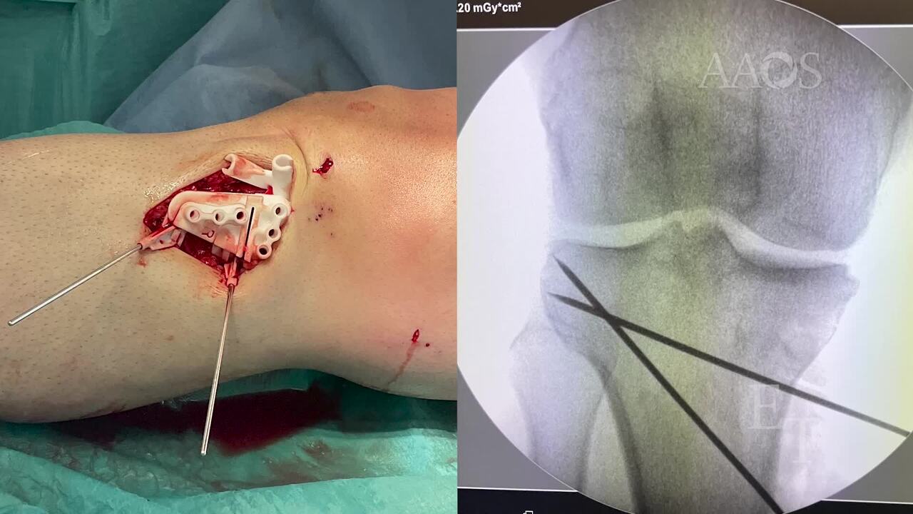
The third step is an opening wedge high tibial osteotomy. The patient-specific instrumentation guide is applied in the same position using the two guide pins left in place. The osteotomy is performed under fluoroscopic guidance. The patient-specific instrumentation guide prevents damage to the tunnels, which are proximal to the upper part of the fixation plate. The medial opening is created to the predetermined height. The patient-specific instrumentation guide is removed. The predefined medial plate is positioned in such a manner that the holes in the plate correspond with the holes already drilled. The plate is fixed proximally with the use of 4.5-mm locking screws and distally with the use of 4.5-mm cortical and locking screws. Allograft bone is used for bone grafting of the osteotomy site.