Excision of Glomus Tumor Using the Nail-Sparing Approach
Glomus tumors are an uncommon occurrence in the hand, accounting for approximately 1% to 5% of all hand tumors. Several approaches to subungual glomus tumors have been described; however, postoperative cosmetic nail deformity is a common complication, particularly if the nail bed is divided during the approach. Given a paucity of surgical videos on glomus tumor excision, the goal of this surgical video is to demonstrate a nail-sparing approach for glomus tumor excision in a step-by-step, reproducible manner.
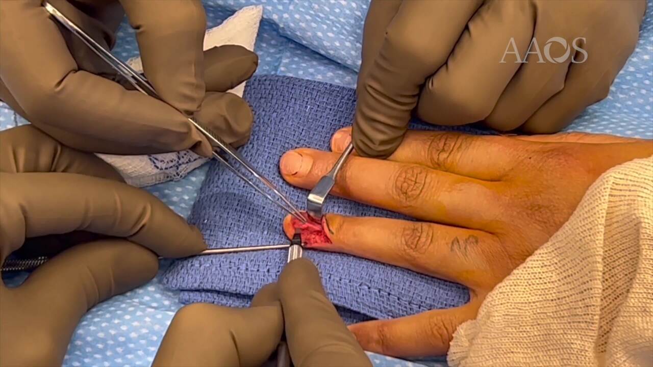
The case presentation of a 43-year-old woman with a painful glomus tumor in her left ring finger is reviewed. The mass was confirmed on MRIs and was confirmed to be the cause of cortical depression of the distal phalanx on radiographs. Given the patient's persistent pain, surgical excision was indicated. An incision was made obliquely on the ulnar eponychial fold. The nail plate was elevated sharply in a proximal to distal direction. The germinal matrix was then elevated from ulnar to radial, exposing the tumor underneath. This technique avoids an incision in the germinal matrix; instead, the germinal and sterile matrix are elevated and placed back down. The tumor was excised, and diagnosis was confirmed by pathology. The germinal matrix was sutured back meticulously with the use of 6.0 plain gut sutures, and the nail was placed back as a stent. The patient was satisfied with the procedure with regard to pain relief, and the cosmetic result was excellent.