Sciatic Nerve and Common Peroneal Nerve Neurolysis
This video describes sciatic nerve and common peroneal nerve neurolysis. The patient described in this video was indicated for sciatic nerve neurolysis after a clinical evaluation and a magnetic resonance neurogram demonstrated scarring about the sciatic nerve and compression of the common peroneal nerve at the level of the fibular head.
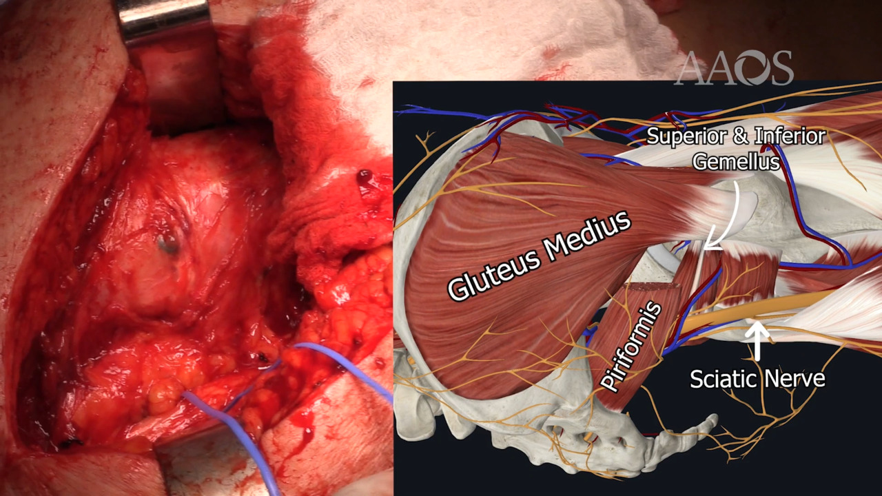
The prior incision is opened and extended distally. Dissection is continued through superficial and subcutaneous tissue until the iliotibial band and the gluteus maximus are identified. Dissection proceeds through ample scar tissue from prior procedures. After the gluteus maximus is reached, it is divided in line with its fibers until the first neurovascular bundle is identified. Scar tissue is dissected off the posterior capsule and the short external rotator repairs, which allows for identification of the piriformis. The piriformis is then divided, allowing for palpation of the sciatic nerve proximally. Dissection is continued to isolate the sciatic nerve and allow for neurolysis. During neurolysis, the scarred epineurium is removed from the nerve. Neurolysis is continued through intraneural scarring, and the nerve is separated into its tibial and common peroneal components. Concurrently, a curvilinear incision is made over the fibular head. Dissection through subcutaneous tissue allows for identification of the common peroneal nerve. The common peroneal nerve is released proximally and distally, and scar tissue is removed.
After neurolysis, nerve stimulation of the tibial and common peroneal distributions of the sciatic nerve is performed. The nerve is protected with the use of fibrin glue, and the wounds are closed in a layered fashion with the use of absorbable suture.