Minimally Invasive Sacroiliac Joint Arthrodesis
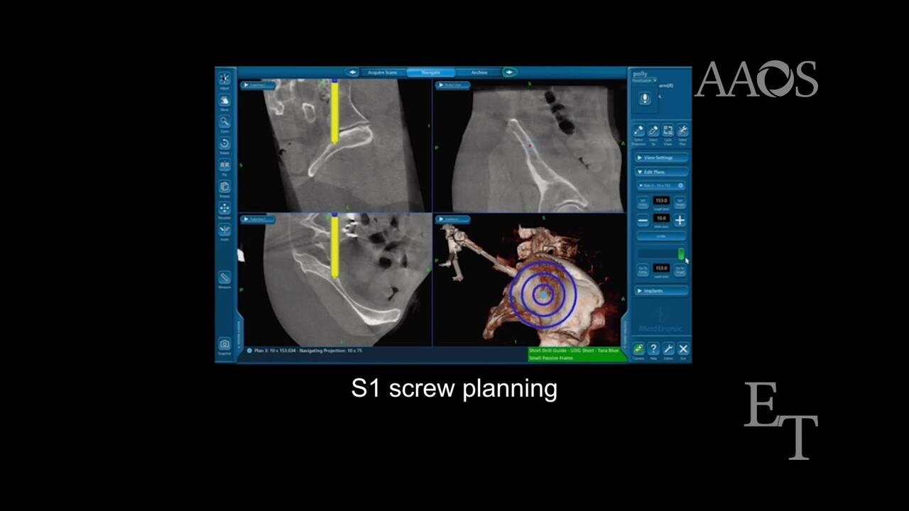
This video demonstrates safe and effective minimally invasive sacroiliac joint arthrodesis via a lateral approach using fully threaded three-dimensionally printed screws and navigation, indicated for sacroiliac joint pain because of degenerative sacroiliitis and/or sacroiliac joint disruption. The case presentation of a patient with symptomatic bilateral sacroiliitis refractory to nonsurgical management, including physical therapy, medications, and local anesthetic and steroid injections, is discussed. Preoperative imaging studies and surgical planning are discussed, and the rationale for surgical treatment is provided.This procedure is performed with the patient under general anesthesia and in the prone position, using three-dimensional navigation, such as cone-beam CT. After navigation setup, including reference pin placement in the contralateral ileum, a navigated probe is used to approximate the desired location, trajectory, and size of each implant. The positions are marked on the skin, and a 3- to 5-cm lateral incision is made. The gluteal fascia is then bluntly dissected to the outer table of the ilium, and soft-tissue protectors are placed. A through-and-through pin is passed across the sacroiliac joint and into the contralateral ilium using three-dimensional navigation. Alternatively, if a through-and-through technique is not used, the pin is passed across the sacroiliac joint and into the center of the sacrum lateral to the neural foramina. After radiographic confirmation of correct pin placement, a hole is drilled using the pin as a guide. Typically, two implants are used for unilateral fusion (one in S1 and one in S2), and four implants are placed for bilateral fusion (one in S1 and one in S2 per side). Postplacement imaging studies of the pelvis are then obtained. Lastly, the wound is irrigated and closed in layers.