Ankle Arthrodesis Through a Modified Scranton Method
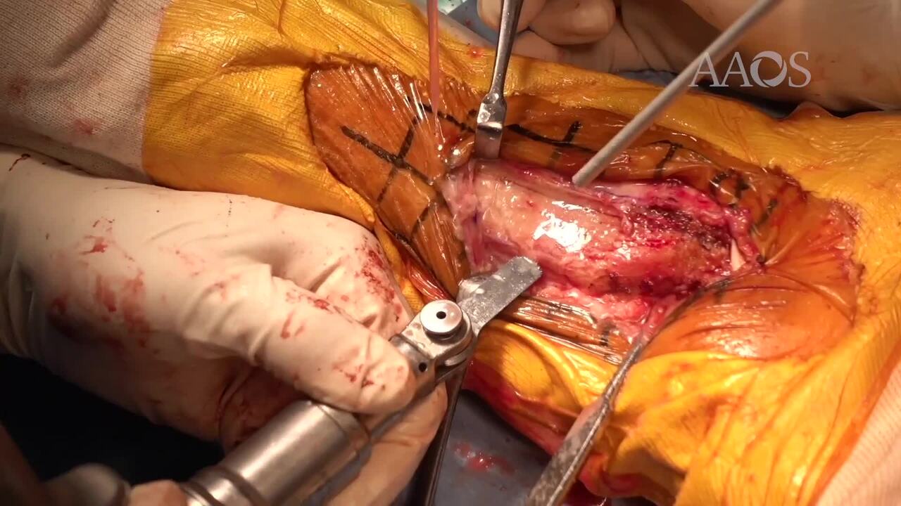
Ankle arthrodesis is a useful treatment option for ankle disorders with limited range of motion. Many studies have been published on various approaches and methods of fixation. The Scranton method was first described in 1985 and involves ankle arthrodesis via resection of the anterior two-thirds of the medial malleolus, bone grafting from resected bone, and fixation with the use of a nonlocking T-plate. This video describes ankle arthrodesis via a modified Scranton method. This modified method does not include a fibular osteotomy, involves the use of one cannulated screw, and involves fixation with the use of a humeral locking plate.
Postoperatively, a cast or splint is not used. Full–weight bearing in an aircast boot is allowed 2 weeks postoperatively. Full–weight bearing without an aircast boot is allowed 10 to 12 weeks postoperatively, depending on the progress of bone union.
The authors of this video managed 39 ankles. The mean age of the patients was 68 years. Using the Takakura-Tanaka classification system, two ankles were classified as stage IIIa, 14 ankles were classified as stage IIIb, 20 ankles were classified as stage IV, and three ankles were not classified. The follow-up period was 33 months. The mean Japanese Society for Surgery of the Foot ankle and hindfoot score improved from 55.6 points to 85.1 points. Complications included a fracture at the proximal end of the plate in one ankle; however, bone fusion was achieved without surgical management or cast immobilization. Nonunion was reported in one ankle; however, no symptoms were reported. No wound complications were reported, and the fusion rate was 97.4%.
The modified Scranton method is associated with some advantages. A fibular osteotomy is not performed, which leads to minimum shortening and preservation of the stable lateral wall. One cannulated screw is used, which affords compression of the arthrodesis site. A humeral locking plate is used, which has sufficient screw holes and is shaped to fit the medial malleolus. These advantages afford solid fixation and early postoperative care.