Custom-Made 3D-Printed Combined Total Talar Replacement Implanted With Patient-Specific Instrumentation (PSI)
Introduction
End-stage ankle joint arthritis is a disabling condition that affects about 1% of the general population, and its incidence is increasing over time, with relevant high social and economic costs.
Historically, the reference standard treatment for ankle arthritis has been ankle fusion, which relieves pain at the cost of tibiotalar motion.
Total ankle arthroplasty (TAA) allows to overcome these issues, and in recent years its indications have been extended. However, some contraindications are still present, such as talar bone loss due to avascular necrosis of the talus, talar disruption, or avulsion.
In this scenario, total talar replacement (TTR) has the potential to overcome much of these limitations. The literature has proven its effectiveness, although it has some issues, such as tibial plafond wear and difficult implant positioning owing to posttraumatic anatomic deformities.
The use of computer-aided surgery and custom cutting guides, supported by computed tomography (CT) scans and by preoperative planning software, have the potential to improve surgical accuracy.
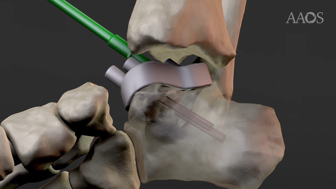
The aim of this video is to present the surgical treatment of nine consecutively treated posttraumatic arthritic ankles with a severe talar collapse by implantation of a custom-made 3D-printed combined TTR implanted with patient-specific instrumentation (PSI).
Materials and Methods
This was a retrospective study. We included all patients who underwent a total talar replacement with usage of PSI, in which TAA were not indicated because of a severe loss of talar bone stock, between January 2017 and December 2019.
All patients underwent a weight-bearing bilateral ankle TC scan. The shape of the required prosthesis was then designed to reconstruct the missing bone to fit the remaining joint surfaces. The custom-built prosthesis was fabricated by “mirroring” the contralateral normal ankle. Polygon manipulation was used to obtain the corresponding bone resections as well as the corresponding PSI, designed to match exactly the frontal bone of the ankle and embed all necessary guides for bone preparation, including the locations of Kirschner wires and levels of bone cuts and drills. All the procedures were performed by the same experienced surgeon.
Patients were evaluated via clinical examinations and radiologic assessments. Mean follow-up was 26.3 months (range, 24 to 36 months). Patient-reported outcome measures questionnaires were submitted preoperatively and at the last follow-up to ascertain patients' satisfaction with the surgical treatment.
Results
A total of 9 patients (6 men and 3 women) were included, with a mean age of 47.7 years (range, 35 to 74 years). Mean follow up was 26.3 months (range, 24 to 36 months).
The mean American Orthopaedic Foot & Ankle Society Score improved from 34.4 ± 5.6 preoperatively to 78.6 ± 6.2 postoperatively. The mean score on the Manchester-Oxford Foot Questionnaire improved from 83.3 ± 14.4 preoperatively to 31.0 ± 15.6 postoperatively. The mean score on the 36-Item Short Form Survey improved from 15.3 ± 4.9 preoperatively to 84.6 ± 5.8 postoperatively.
Mean ankle dorsiflexion improved from 5.2° ± 1.9° preoperatively to 13.1° ± 2.8° postoperatively. Mean ankle plantar flexion improved from 7.4° ± 1.6° preoperatively to 25.7° ± 5.5° postoperatively. No signs of implant mobilization were detected at the radiologic follow-up. No major complications were reported.
Discussion
Although TTR has been already described in the literature, implant availability, costs of production, and surgical reproducibility are still unsatisfactory.
Custom-made 3D-printed combined total talar replacement has proven effective in both restoring ankle motion and relieving pain. The use of PSI allowed a more accurate implant positioning, short operative time, low fluoroscopy exposure, and, theoretically, decreased perioperative complications.
Conclusions
Custom-made 3D-printed combined total talar replacement implanted with patient-specific instrumentation is a valid surgical option in case of severe talar bone loss, restoring ankle motion and relieving pain. Despite promising results, evaluation of the long-term outcomes in a higher number of patients is needed.