Customized Osteotomy of the Radius and Ulna for Longstanding Deformity: The Cutting Edge
Case Overview: We present the case of a 27-year-old man with chronic wrist pain and limited forearm rotation secondary to chronic malunion of the radial and ulnar shafts.
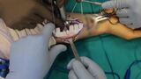
Technique: Before surgical intervention, the patient underwent bilateral CT scans of the forearms for the fabrication of custom specific jigs. The patient was positioned supine with a standard hand table. An 8-cm incision was made over the volar radius by using the approach of Henry. On completion of the deep dissection, the guides were installed, and its placement compared to the three-dimensional (3D) model, with Kirschner wires applied to fix its position. Guide holes were drilled with a 2.5-mm bit, and appropriate cuts were made along the guide. This was then completed for the ulna via the subcutaneous approach. The plates were then bent according to our 3D model and fixed to the ulna and radius. A motion check was performed and revealed full pronation and supination without hindrance.
Results: This patient demonstrated excellent results. At 2 weeks after surgery, the patient had full pronosupination of the forearm with near minimal pain. Correction of malunited forearm fractures via computer-assisted osteotomy, described by Murase et al in 2008 and by Miyake et al in 2012, has been shown to greatly improve pain and clinical range of motion even in patients with long-standing deformities.
Summary: The use of a customized 3D model for corrective osteotomy is a viable treatment option for patients with chronic pain and functional deficiency secondary to malunited forearm fractures, even in patients with longstanding deformity of more than 10 years.