Arthroscopically Assisted Posterior Glenoid Reconstruction With Distal Tibia Osteochondral Allograft
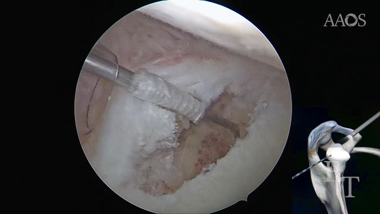
Introduction Posterior glenoid bone loss occurs in more than two-thirds of patients with posterior glenohumeral instability, with 28% having greater than subcritical bone loss (13.5%), a marker for potential need for bony augmentation versus soft tissue-only procedures. Several techniques are described to augment either the version or volume of the glenoid surface including osteotomies, autograft transfers, and allograft tibia transfers. Indications Arthroscopically assisted allograft distal tibia bone block augmentation to the posterior glenoid is indicated for revision posterior instability procedures with posterior bone loss and in primary cases of posterior instability with critical bone loss. Technique Our technique for arthroscopic posterior glenoid reconstruction with allograft distal tibia and posterior labral repair is performed in the lateral position, involves the use of standard instrument sets, and requires no patient repositioning. The planned tibial bone block is prepared on a back table either prior to, or concurrently with, the arthroscopic procedure. After creation of high posterior and standard anterior portals, a sucker-shaver and burr are used to create a perpendicular edge for apposition of the allograft tibia. The bone block is introduced through a longitudinal incision and underdelivered to the prepared surface under the liberated labrum. The articular surface of the graft and glenoid are aligned, and cannulated screws are used to compress the bone block against the native glenoid. The posterior labral tissue is then mobilized over the graft and repaired to the native glenoid. Discussion and Conclusion The benefits of allograft tibia augmentation for posterior instability with glenoid bone loss include an anatomic joint surface restoration including articular cartilage, lack of donor site morbidity, and a minimally invasive approach. When performed arthroscopically, this technique permits concurrent posterior labral repair and anatomic reconstruction.