Bicortical Titanium Tenodesis Button With Double-Row Fixation for the Management of Proximal Rectus Femoris Avulsion
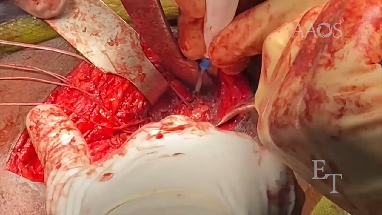
Introduction Rectus femoris avulsions are reported in adult professional soccer and football players but are exceptionally rare. No preferred treatment method or treatment recommendations exist for the management of rectus femoris avulsions; however, in patients with an anterior inferior iliac spine avulsion, rest, immobilization, and rehabilitation appear to result in positive outcomes. Typically, surgical treatment is reserved for patients with large retractions of bone fragments and patients in whom nonsurgical treatment is unsuccessful. Surgical treatment options include direct suture repair, single- and double-row bone suture repair, and muscle-muscle repair. This video presents our technique for the management of a severely retracted rectus femoris tendon avulsion in a high-level athlete, which involves the use of a bicortical tenodesis button and double-row fixation. Case Presentation The case presentation of a 24-year-old professional soccer player with severe pain in the anterior hip after a deceleration injury and a fall during a soccer match is discussed. The patient’s physical examination revealed tenderness to palpation just distal to the anterior inferior iliac spine, pain and weakness with hip flexion, and inability to perform a straight leg raise. MRI of the left hip revealed a complete avulsion of the rectus femoris tendon with 5 cm of retraction. Surgical Technique Intraoperative ultrasonography was used to localize a 6-cm incision created following the Smith-Petersen approach between the tensor fascia latae and the sartorius. The rectus femoris tendon was released of adhesions and surrounding tissue. The rectus femoris footprint on the anterior inferior iliac spine was prepared down to bleeding subchondral bone with the use of a high-speed burr. The tendon was prepared via a locking Krackow stitch technique, and sutures were placed in a titanium tenodesis button that was drilled and inserted bicortically on the inferior aspect of the anterior inferior iliac spine. A SwiveLock anchor (Arthrex) loaded with sutures from the cortical button and independent nonabsorbable sutures were placed above the tenodesis button on the superior aspect of the anterior inferior iliac spine. The anchor islet sutures were then placed in the tendon in a locking fashion and secured with the use of a second superior row anchor for increased surface area compression of the tendon on the footprint. Postoperative Rehabilitation The patient was instructed to remain toe-touch weight bearing for 2 weeks postoperatively, with blood flow restrictions to the surgical leg. At 4 weeks postoperatively, the patient began physical therapy for proprioceptive exercises, range of motion, and single-leg squats for strengthening. At 6 weeks postoperatively, the patient started soccer-specific drills and light jogging. The patient fully returned to professional soccer at 8 weeks postoperatively. Conclusion This video presents our technique that involves the use of a bicortical tenodesis button and double-row fixation for the management of a severely retracted rectus femoris tendon avulsion in an elite athlete. The technique resulted in successful accelerated return to professional soccer.