Arthroscopic Fixation of Os Acetabuli/Acetabular Rim Fracture
Background
Resection of a large segment of the acetabular rim may result in hip instability or increased progression of osteoarthritis in the joint. Internal fixation of os acetabuli (OSA)/rim fracture (RF) is indicated if the lateral center edge angle is less than 25° on an AP radiograph or the anterior center edge angle is less than 20° on a false-profile radiograph after removal of the osseous fragment. Arthroscopic fixation of acetabular rim fractures was first described in 2009. The incidence of OSA or RF in patients undergoing hip arthroscopy for the management of femoroacetabular impingement (FAI) is between 3.5% to 7.7%. OSA/RF typically is observed in males and usually is associated with CAM morphology. The terminology of OSA and RF often overlap. OSA and RF are differentiated by pathogenesis and radiographic appearance. OSA is of cartilaginous origin as an unfused ossification center and appears parallel to the joint surface. RF is a result of a repetitive loading of the acetabular rim by a CAM lesion, leading to a stress fracture that is oriented perpendicular to the hip joint.
Purpose
This video overview and case presentation demonstrates a technique for arthroscopic acetabular rim fracture fixation in a patient with FAI and a labral tear.
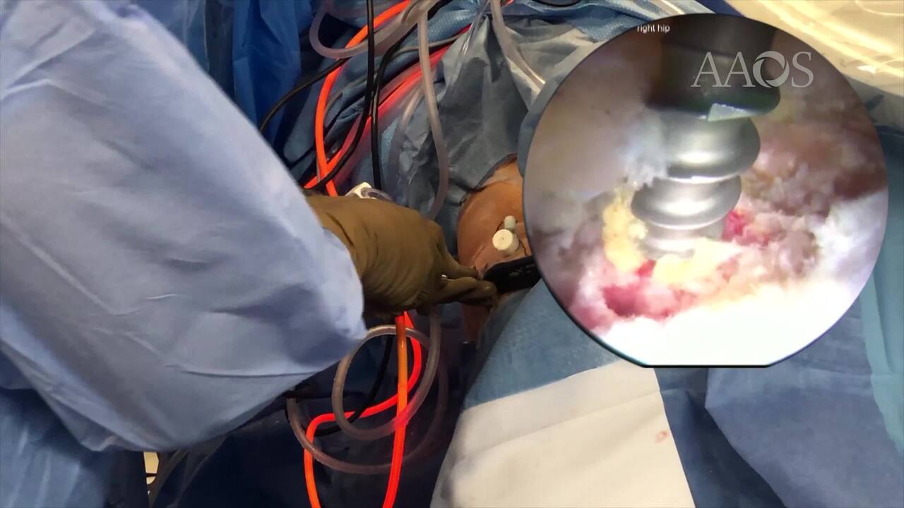
Methods
An overview of the pathogenesis, diagnosis, and treatment of a patient with FAI and an acetabular rim fracture is provided. The case presentation of a 24-year-old man with right hip FAI and an acetabular rim fracture is discussed. A surgical technique for arthroscopic acetabular rim fracture fixation and labral repair with femoral neck osteochondroplasty is demonstrated.
Results
Initial postoperative radiographs demonstrated that the procedure restored the acetabular rim and resected the CAM lesion on the femoral neck. The patient is progressing toward his goal of returning to marital arts.
Conclusion
Arthroscopic fixation of an acetabular rim fracture is indicated if removal of the osseous fragment may lead to instability or progression of osteoarthritis. Early results suggest patients can achieve excellent radiographic and clinical results.