Capsule-Preserving Approach to Arthroscopic Decompression of the Subspine
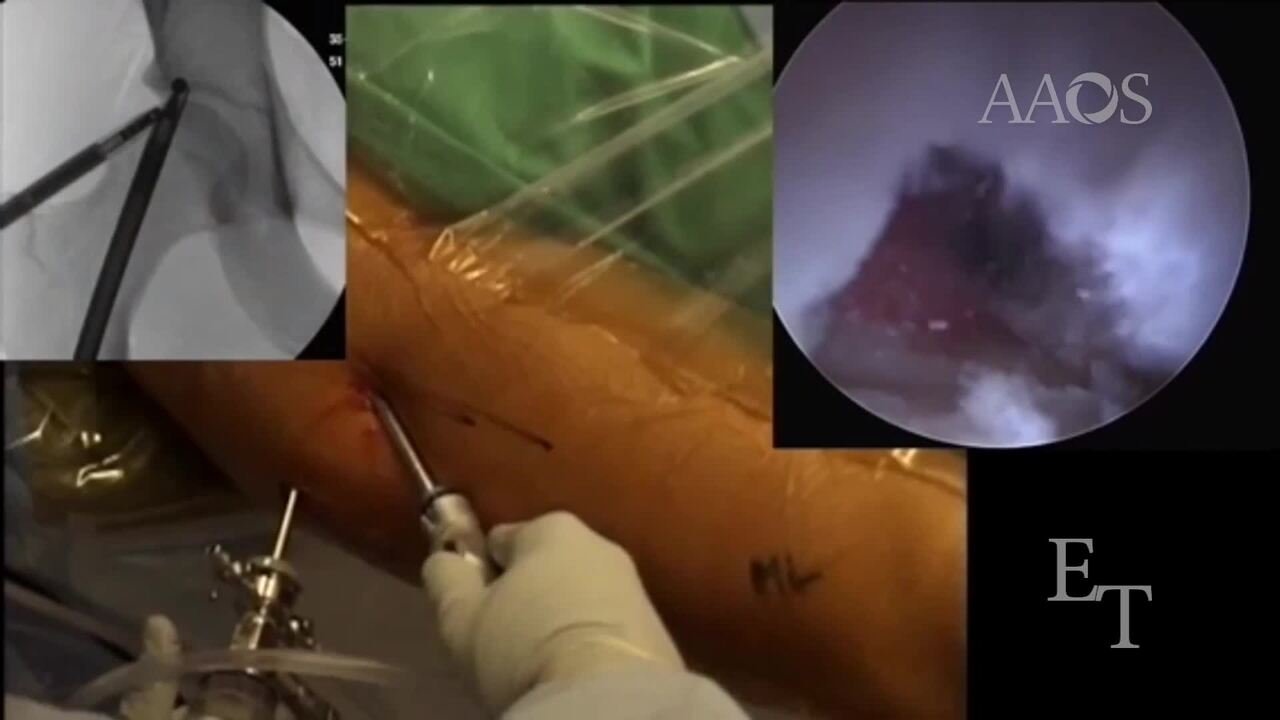
After standard diagnostic arthroscopy, all intra-articular pathology, including labral tears and chondral flaps, should be managed. Immediately above the anterosuperior labrum, between the 1-o'clock and 3-o'clock positions, is the most common area of anterior inferior iliac spine (subspine) impingement; this should be correlated with fluoroscopic images and three-dimensional CT scans. The base of the subspine at the level of the joint can be managed from within the interportal capsulotomy; however, to manage the entire subspine deformity without creating an inverted L-shaped capsulotomy, a pericapsular window must be created. A banana-style beaver blade is slotted into the joint, and a new path through the rectus and the proximal capsule is created under direct visualization. Through the path that was created with the use of the beaver blade, the slotted canula and the switching stick are placed. Then, the burr is taken apart, and the sheath is placed over the switching stick followed by the inner burr component. Fluoroscopy is used to confirm adequate subspine resection through this window. Adequate decompression is confirmed via a false-profile view by shifting the fluoroscopic image intensifier back 40°. Occasionally, for avulsion-type or large deformities, two or three pericapsular windows are necessary to complete the subspine resection. Subspine impingement should be assessed and managed in patients with femoral anteversion of less than 5° and in patients with a type II or type III anterior inferior iliac spine deformity. These small pericapsular windows preserve the proximal capsule and the interportal capsulotomy and allow for adequate capsular closure without risking instability. The femoral head-neck offset or cam deformity can then be corrected through the interportal capsulotomy. Our preference is to close the interportal capsulotomy with the use of four to six simple, nonabsorbable stitches after comprehensive femoroacetabular impingement correction.