Management of Severe Ankle and Hindfoot Deformity: Technique Using Femoral Head Allograft for Tibiotalocalcaneal Fusion Using a Cup-and-Cone Reamer
Introduction
Limb shortening as a result of structural bone loss in patients with tibiotalocalcaneal arthrodesis may negatively affect gait and weightbearing. The use of a femoral head allograft and an intramedullary nail during tibiotalocalcaneal arthrodesis successfully preserves limb length in patients with structural bone deficits. This video presents a technique involving the use of a femoral head allograft and a cup-and-cone reamer for the management of severe ankle and hindfoot deformity.
Case Presentation
A 64-year-old patient with a severe, fixed ankle valgus deformity underwent ankle arthrodesis via a lateral approach and a fibular osteotomy. At 1 year postoperatively, the patient had incomplete deformity correction and a nonunion with a potentially neuropathic joint. The patient underwent hardware removal, bony exostectomy, a fibular osteotomy of the distal portion of the fibula, irrigation and debridement, biopsy of the soft tissues of the foot and ankle, and placement of a cement spacer.
Surgical Technique
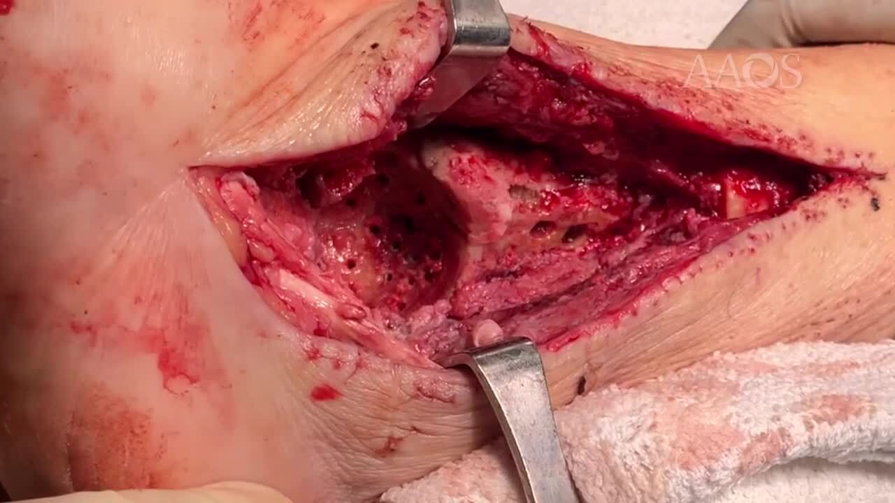
The distal tibia and the remaining talus or calcaneus are prepared with the use of an acetabular reamer, which creates a concave surface for secure placement for the interposition of a femoral head allograft. This increases the amount of bone-to-bone contact between the allograft and the residual host bone, providing a stable construct for alignment and placement of the intramedullary nail for tibiotalocalcaneal fusion.
Conclusion
Femoral head allograft for tibiotalocalcaneal fusion may be useful in patients with a large talar body defect and a severe fixed deformity of the ankle and hindfoot.