High Thoracic Pedicle Subtraction Osteotomy in the Management of Severe Congenital Kyphoscoliosis in the Adult
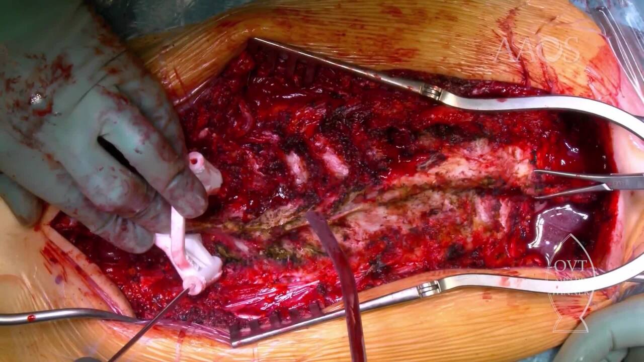
Introduction Congenital kyphoscoliosis is a three-dimensional pathology that results from abnormal embryonic development of the spine vertebrae. According to the McMaster classification, principal causes of congenital kyphoscoliosis include failed formation of the vertebra (type I), failed segmentation (type II), and failed formation of the vertebra and failed segmentation (type III). Congenital kyphoscoliosis in adults, if unmanaged, may lead to invalidating clinical conditions. In patients with a highly rigid, severe spinal deformity, vertebral osteotomies via a posterior approach with the use of pedicle screw instrumentation only effectively achieves spinal balance, stability, and correct alignment. Particularly, pedicle subtraction osteotomy (PSO) involves removing all the posterior elements of the vertebra (ie, spinous process, laminae, pedicles, and facets) to gain better access to the vertebral body, the location at which a wedge osteotomy is performed. Closure occurs in a wedge fashion and allows for kyphosis correction via shortening of the posterior column. A PSO affords 30&[deg] to 40&[deg] of lordotic correction at each level, which is suitable for patients with highly rigid kyphosis. If coronal correction is necessary, then an asymmetric PSO can be performed. Materials and Methods We performed a retrospective study of all patients who underwent posterior spine correction and fusion for severe congenital kyphoscoliosis (thoracic kyphosis >70&[deg]) at our institution between January 2015 and June 2018. All the patients underwent single-stage posterior arthrodesis with the use of high-density pedicle screws. All the patients underwent low-dose, high-resolution CT of the spine. A virtual model was created, and the entry points and the trajectories of the screws and the main geometric parameters were set by the surgeon. Customized drill guides were printed accordingly and used for screw placement. Sensory- and motor-evoked potentials were monitored. In this case series, all the patients underwent a high thoracic PSO. All the procedures were performed by the same experienced surgeon. All the patients underwent postoperative CT to assess the trajectories of the screws and to compare them with CT scans used for preoperative planning. The trajectory of all the screws was then evaluated according to the Gertzbein-Robbins classification. All the radiographic measurements, which were obtained to assess deformity correction were obtained by the same experienced surgeons who performed the surgical procedures. The mean follow-up was 2.38 years (range, 2 to 2.7 years). A Scoliosis Research Society Outcomes 22-Item Questionnaire was submitted preoperatively and at final follow-up to determine patient satisfaction with surgical treatment. Results A total of five patients (four females, one male) with a mean age of 32.2 years (range, 20 to 49 years) were included in the study. The mean surgical time was 263 minutes. Mean thoracic kyphosis improved from 74.7&[deg] &[plusmn] 13.1&[deg] to 42.7&[deg] &[plusmn] 11&[deg], resulting in a correction rate of 42.8% &[plusmn] 5.2%. The mean Cobb angle decreased from 41.2&[deg] &[plusmn] 12.4&[deg] to 27.3&[deg] &[plusmn] 8.9&[deg], resulting in a correction rate of 33.74% &[plusmn] 6%. The patients were satisfied with the outcome of the procedure, and the mean Scoliosis Research Society Outcomes 22-Item Questionnaire score increased from 2.1 &[plusmn] 0.4 preoperatively to 3.3 &[plusmn] 0.5 postoperatively. No major intraoperative or postoperative complications were reported. Postoperative CT scans showed highly accurate screw trajectories that were completely superimposable with those on CT scans used for preoperative planning. The cortex was violated only if violation was planned to optimize screw strength. According to the Gertzbein-Robbins classification, 91.3% of the screws were completely inside the pedicle and 8.7% were grade A; zero were grade B or grade C. Discussion and Conclusions Congenital kyphoscoliosis may result in sagittal imbalance, pain, neurologic complications, progression of deformity, cardiopulmonary disease, or interference with activities of daily living. Vertebral osteotomies may be the only therapeutic method to properly correct the deformity. The literature reports that vertebral osteotomies are very effective but may be complicated by bleeding, neurologic damage, and dural lesions. Although a technically demanding procedure, thoracic PSO via a posterior approach with the use of pedicle screw instrumentation only is an effective and safe procedure for the correction of highly rigid, severe spine deformities, such as congenital kyphoscoliosis, in adults if performed by an experienced surgeon. The use of patient-specific drill guides may aid in screw placement in patients with highly dysplastic pedicles, decreasing the risk of intraoperative and postoperative complications.