Arthroscopic Débridement of Elbow Radiocapitellar Plica
Elbow synovial plica syndrome is an uncommon cause of elbow pain. Synovial folds may become inflamed and hypertrophic, resulting in mechanical symptoms with range of motion of the radiocapitellar joint. The clinical presentation of elbow synovial plica syndrome may be vague, and elbow synovial plica syndrome often is mistaken for lateral epicondylitis because patients are tender in similar anatomic areas. Patients may report mechanical symptoms on the lateral side of the elbow, such as snapping or popping, which can be elicited via various maneuvers. Anterior plica snapping can be reproduced via elbow flexion with pronation, and posterior plica snapping can be reproduced via elbow extension with supination.
Plain radiographs are obtained to rule out any other pathology. Ultrasonography may reveal enlargement of the synovial plica and can be used to dynamically visualize tissue snapping in the radiocapitellar joint. MRI can be obtained to visualize hypertrophic radiocapitellar plica.
First-line nonsurgical treatment consists of physical therapy and NSAIDs. However, patients with mechanical symptoms and snapping may benefit from arthroscopic surgical débridement of the plica tissue. Surgical treatment is indicated in patients with persistent pain and mechanical symptoms. A few case series have reported excellent outcomes after arthroscopic débridement of radiocapitellar plica, which results in minimal to no complications.
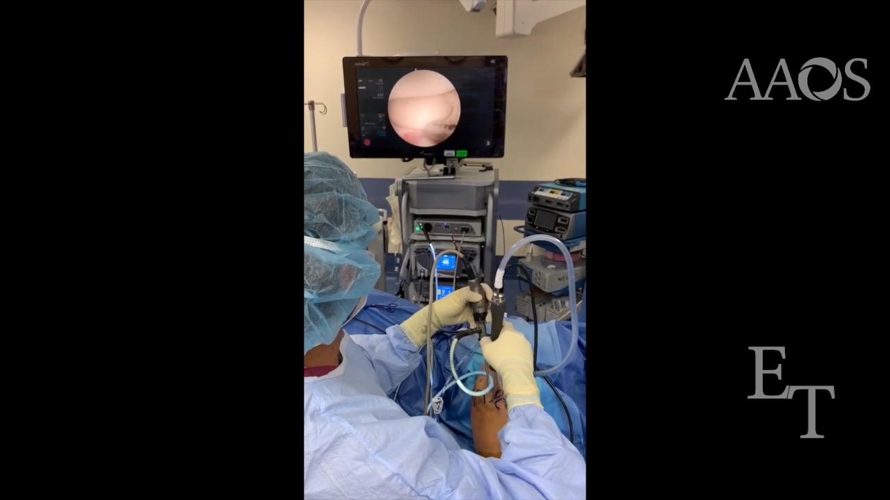
This video demonstrates the surgical technique for arthroscopic débridement of radiocapitellar plica in a patient with lateral elbow pain and snapping. Relevant case series, patient presentation, physical examination findings, imaging studies, technical pearls of the procedure, and postoperative rehabilitation protocol are reviewed.