Use of a Carbon Fiber Rod Augment During Nonarticulating Antibiotic Knee Spacer Placement
Chronic prosthetic total knee infections typically are managed via two-stage revision knee arthroplasty. The surgical technique involves explantation of the infected prosthesis, placement of an antibiotic cement spacer, removal of the spacer, and revision knee arthroplasty. In the described technique, the authors of this video advocate the use of an intramedullary carbon fiber rod augment during the placement of a nonarticulating antibiotic spacer to enhance stability, prevent bone loss, and promote soft-tissue healing in patients with extensor mechanism deficiency or local soft-tissue compromise.
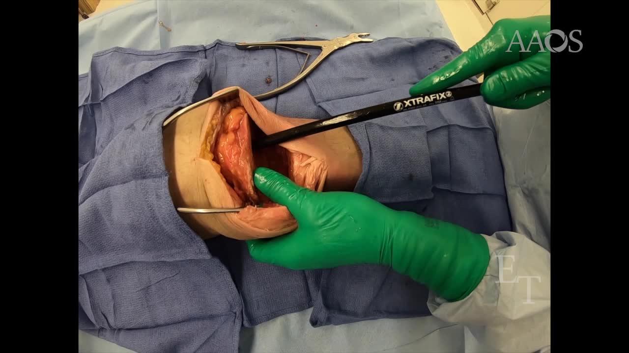
During the first stage of the procedure, a standard medial parapatellar approach is used, and the infected prosthesis is explanted. An intramedullary rod is then inserted in the femoral and tibial canals, centered in the knee joint. A guidewire and flexible reamer may be used to prepare the intramedullary canal for rod placement, if necessary. A standard nonarticulating antibiotic cement spacer is placed around the intramedullary rod. To decrease the risk of thermal injury to the posterior neurovascular structures as the cement hardens, the surgeon may elect to place a malleable retractor in the posterior aspect of the joint and/or use continuous irrigation.
After successful management of the infected joint, the antibiotic spacer and carbon fiber rod are removed, and revision knee arthroplasty is performed. First, the cement spacer is fragmented with the use of an osteotome and a mallet. The cement pieces are removed, exposing the carbon fiber rod. The rod is then cut with the use of an oscillating saw and removed with the use of a rongeur or Kocher forceps. Before cutting the rod, sterile ultrasonography gel is placed around the location of the cut to collect carbon fiber debris. The joint is then thoroughly irrigated, and revision knee arthroplasty is performed.
We have used this technique at our institution in a cohort of eight patients, three of whom had quadriceps tendon defects and four of whom had bone loss. No subsequent infections occurred during a follow-up that ranged from 6 to 29 months. All the patients with quadriceps tendon defects had functional extensor mechanism function after primary repair and carbon fiber rod use. Of those with quadriceps tendon defects, one had a 30° extensor tendon lag, and two had full extension with no lag. Two complications occurred in the cohort. The distal end of the rod led to an anterior tibial cortical defect in one patient, who required a long-stemmed tibial component at the time of revision knee arthroplasty to bypass the anterior cortical defect. One patient was discharged to hospice and died from complications of sepsis.
We have found that the use of an intramedullary carbon fiber rod augment is beneficial in select patients with a prosthetic joint infection who require staged revision knee arthroplasty. The outcomes of the eight patients in our cohort are encouraging; however, additional research is necessary to improve and expand this novel technique.