Surgical Anatomy of Segond Fractures
In 1879, Paul Segond conducted a series of cadaver model studies and described a relatively constant avulsion-type fracture in the anterolateral aspect of the tibial plateau caused by forced internal rotation of the knee. This injury was termed a Segond fracture and considered a pathognomonic sign of an anterior cruciate ligament (ACL) tear. Segond reported that a pearly, resistant fibrous band located in the anatomic position of this avulsion injury became tight with internal rotation of the knee. This anatomic structure has been described in various anatomic studies and given various names, such as the middle one-third of the lateral capsule, the anterolateral femorotibial ligament, the anterolateral ligament, and the anterolateral complex. Segond fractures are a subject of interest with regard to the ongoing debate on the soft-tissue structures in the lateral aspect of the knee.
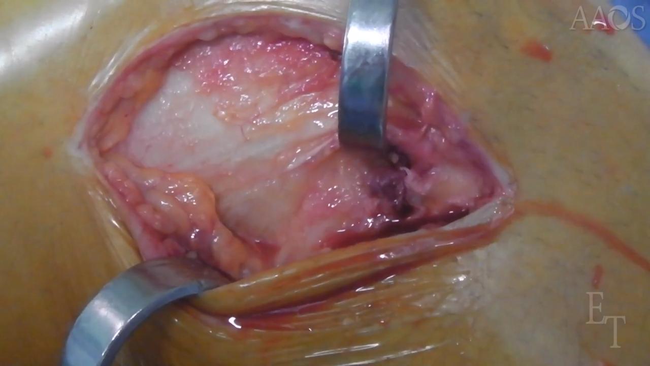
This video investigates the relation of Segond fractures to the anterolateral complex of the knee. We hypothesized that the tibial anterolateral capsule in the knee would match the described location of Segond fractures. The study included 128 consecutive patients who underwent reconstruction for the management of an acute ACL rupture between January 2013 and December 2016. Inclusion criteria were an acute ACL tear that was surgically managed within 10 days postinjury and a Segond fracture observed on radiographs and MRI. At the time of ACL reconstruction, the lateral compartment was exposed in all the patients via a hockey-stick incision. The fascia lata was inspected and longitudinally divided along its fibers to expose the lateral compartment from the posterolateral corner to the Gerdy tubercle. All Segond fractures found were recorded and photographed. All the Segond fractures were directly repaired via a technique based on the dimension of the bone fragment (direct suture, anchors, or screw fixation). The surgical knee was placed in a brace in full extension for 4 weeks postoperatively. Patients were permitted to immediately bear weight as tolerated with the brace and crutches. Progressive range of motion exercises were initiated after the first postoperative day. Daily isometric and isotonic exercises were prescribed. At 4 weeks postoperatively, full weight-bearing without crutches or the brace was permitted. At 8 weeks postoperatively, a muscle-strengthening program with open kinetic chain exercises was initiated. At 4 months postoperatively, a 1-month gradual return to noncontact athletic and sport-specific training, including running and cutting, was encouraged. Return to full sports activity was allowed at 5 months postoperatively based on an evaluation and a determination of readiness for return to sports activity. Of the 128 patients, 89 were men and 39 were women. A Segond fracture was observed on the preoperative radiographs of 12 of the patients (9.3%). The Segond fractures were repaired via periosteal stitches in seven patients, via suture anchors in four patients, and via a cancellous screw in one patient because of a larger fragment. Surgical exploration of the anterolateral knee did not reveal injury to the iliotibial band, which appeared to be only mildly stretched or hemorrhagic. A longitudinal incision of the superficial and deep iliotibial band was made, and the Segond fracture was identified. Careful dissection of the Segond fracture revealed a discernible attachment of the anterolateral capsule to the bony avulsion in all of the patients. The bony fragment was not attached to fibers of iliotibial band in any of the patients. The fragment was only attached to the anterolateral capsule. No complications such as infection, malunion of the avulsion fracture, postoperative stiffness, or ACL rerupture were reported. Patients returned to sports activity after a mean time of 6 months. At the latest follow up, all patients had returned to their preoperative sports activity level. In general, Segond fractures are described as an avulsion-type fracture, indicating the detachment of a bone fragment by the pull of a ligament or tendon from its insertion point. Although Segond reported a fibrous band attached to his eponymous fracture, subsequent studies have remained unclear on what structure causes the avulsion.
The research of Dr. Steven Claes supported the hypothesis that the anterolateral ligament inserts in the region of the proximal tibia, the location from which Segond fractures consistently avulse. This suggests that Segond fractures are a bony avulsion of the anterolateral ligament. In a surgical examination of a Segond fracture, Dr. Marcio Albers demonstrated that the posterior fibers of the iliotibial band and the lateral capsule were linked to the bone fragment. This case series supports our hypothesis that the tibial anterolateral capsule inserts on all Segond fractures. Careful dissection of the Segond fractures revealed the discernible attachment of the anterolateral capsule to the bone injury in all the patients.