Pemberton Pericapsular Osteotomy for Developmental Dysplasia of the Hip and Hip Dislocation
2018 HONORABLE MENTION
Pemberton pericapsular osteotomy is a common procedure that was introduced in 1965 for the management of a deficient acetabulum in patients with developmental dysplasia of the hip, which is the most common congenital deformity requiring surgical correction osteotomy to prevent early onset of secondary hip arthrosis. Similar to Dega osteotomy and Salter osteotomy, Pemberton pericapsular osteotomy is an incomplete osteotomy that is widely used in children older than 18 months and younger than 8 years; however, it also is successfully used in older patients (younger than 14 years) and patients with neuromuscular disorders. Pemberton developed this procedure to address two problems. First, the acetabulum is not only shallow but also faces forward and laterally. Second, in a hip dislocation, the femoral head usually is small in relation to the acetabulum, whereas a subluxating hip is large in relation to the femoral head.
The senior author has performed this procedure for approximately 40 years in otherwise normal patients with developmental dysplasia of the hip and patients with neuromuscular disorders, achieving satisfactory results. The approach is not difficult; however, caution is necessary to prevent complications, such as excessive postoperative bleeding, lateral femoral cutaneous nerve lesions, and belt line pain. The technique is fairly simple, affords good options for customizing the cut, and is associated with few complications.
Advantages of the Pemberton pericapsular osteotomy make it a very good treatment option. It is feasible to perform bilateral procedures in one surgical session, pin fixation is not necessary, and a greater capacity for correction exists. In addition, the procedure is associated with a small learning curve. Disadvantages of Pemberton pericapsular osteotomy include alteration in the shape of the acetabulum, which requires the procedure to be performed in early childhood to allow for sufficient time for remodeling to create congruity with the femoral head. The results reported in the literature are excellent, and the results of our series also are satisfactory.
This video reports the results of patients who underwent Pemberton pericapsular osteotomy in the last 10 years, with a mean follow-up of 4.5 years. All data reports have been fully digitized, and radiographs were performed in the same standard manner to reduce bias during evaluation. In the past 15 years, newborn screenings and the increased use of early casting, bracing, and open reduction has reduced the number of patients who require osteotomy. However, we believe it is important for orthopaedic surgeons to understand how to perform this procedure.
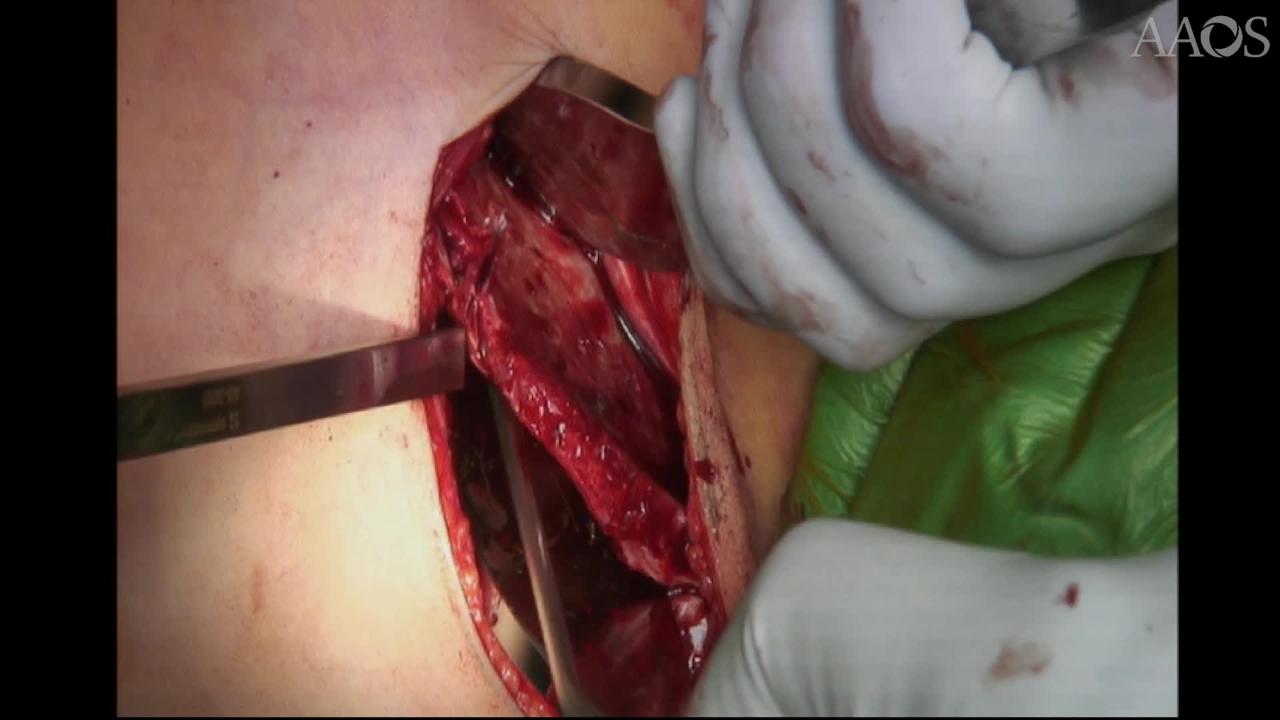
A small incision (bikini incision) is made just distal and parallel to the anterosuperior spine. A hemostat is then used to separate the tissues. The lateral femoral cutaneous nerve is clearly visualized, and the interval distal to the lateral femoral cutaneous nerve is opened very carefully, protecting the nerve. Army-Navy retractors are placed in the interval between the iliotibial band and the sartorius. A Cobb elevator is used to expose the inferior spine. The iliac apophysis is divided sharply to the anterosuperior spine and then along the bridge between the anterosuperior and anteroinferior spine. Gauze is useful for tamponing bleeding and facilitating subperiosteal dissection; it also can be used to protect the soft tissues during dissection. Cobra retractors are used to expose the field. The osteotomy can now be performed. Fluoroscopy is useful to determine the appropriate osteotomy cut if more lateral or anterior coverage is desired. After the cut is determined, the osteotomy can begin. The cut should begin approximately 1 cm above the anterior inferior iliac spine and proceed posteriorly, maintaining a distance of approximately 1 cm to 1.5 cm from the attachment of the joint capsule. The video shows the steps for a safe and satisfactory osteotomy.
After the osteotomy is complete, the acetabular roof can be levered down into the desired position. The cut osteotomy should be opened slowly to allow time for the tissues to follow the spreading force and to avoid fracture. Fluoroscopy can be used to verify that the desired coverage has been achieved. The osteotomy is held open with the spreader, and the graft is placed under compression. A cast is applied with the hip in approximately 30° of flexion, 30° of abduction, and neutral rotation. The cast is removed after 6 weeks.
