Diagnosing Benign Bone-Forming Tumors: A Visual Guideline from Presentation through Treatment
The most common benign bone-forming tumors are enostosis (bone island), osteoma, osteoid osteoma, and osteoblastoma. The presence or production of bone, or osteoid, with growth differentiates benign bone-forming tumors from other bone tumors. Radiographically, benign bone-forming tumors are observed as mineralization, which indicates the presence or production of bone. Clinically, benign bone-forming tumors vary in presentation and biologic behavior. Although enostosis may be asymptomatic and an incidental finding, osteoid osteoma may cause severe pain, especially at night. These entities are unique in their presentation and biologic behavior. For example, osteoblastoma may be locally aggressive and difficult to differentiate from malignant bone-forming tumors. Management ranges from observation to wide resection, depending on the natural history of the tumor and its location. Chemotherapy and radiotherapy are rarely, if ever, used to manage benign bone-forming lesions.
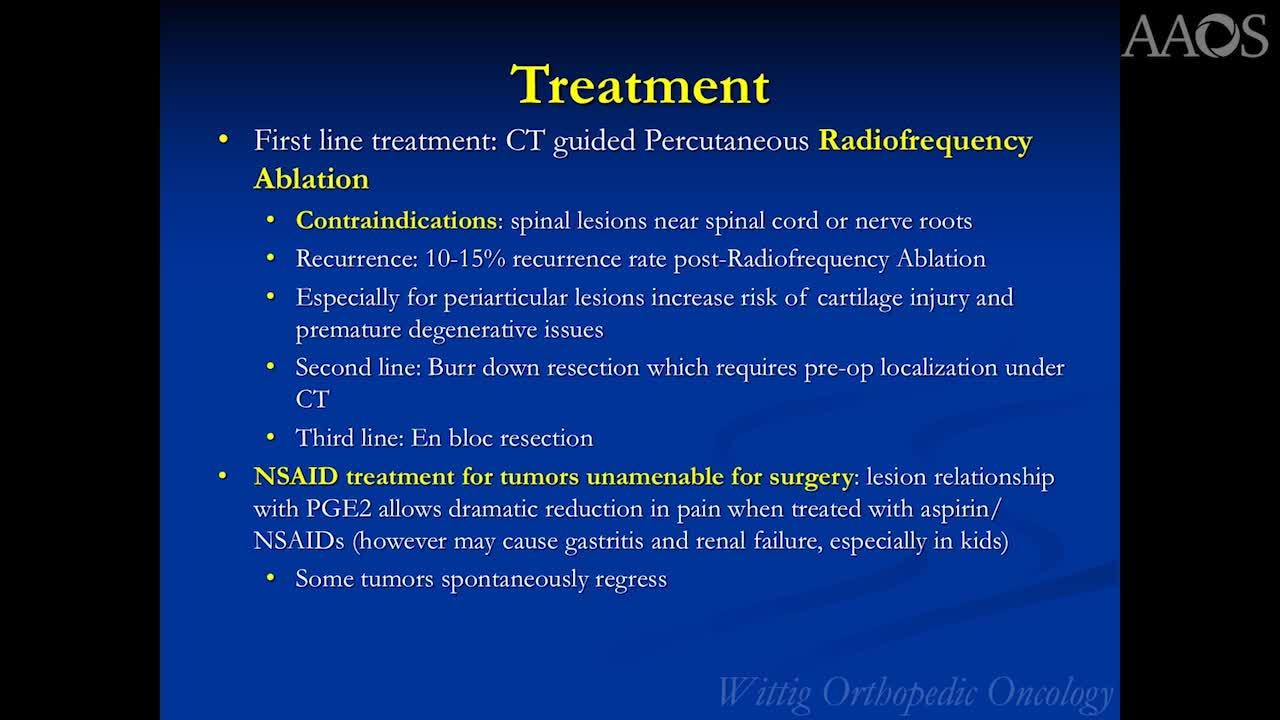
The four aforementioned benign bone-forming tumors can be further divided based on similarities. Histologically, enostosis resemblances osteoma, and osteoid osteoma and osteoblastoma are similar to osteoblastoma. Enostosis and osteoma demonstrate mature cortical or lamellar bone but develop in different locations. Enostosis arises in the medullary canal of the affected bone, whereas osteoma grows on the surface of the bone. Radiographically, enostosis may appear similar to osteoid osteoma. Osteoid osteoma and osteoblastoma are notable for their osteogenic growth of immature woven bone or osteoid arrangement as interlacing trabeculae. However, osteoid osteoma is a self-limiting lesion that rarely grows after presentation. Osteoblastoma is a benign, aggressive tumor that destroys bone as it grows and often has a propensity for local recurrence. Therefore, more aggressive management, including resection of the tumor and possible use of local adjuvants to prevent local recurrence, is necessary for osteoblastoma. Microscopically differentiating between osteoblastoma and osteosarcoma may be difficult, especially without radiographic correlation. Patients should be referred to an experienced orthopaedic oncologist for proper biopsy and treatment. A diagnosis should be rendered only after close correlation with plain radiographs and other pertinent imaging studies.