Implant Removal In Revision Total Knee Arthroplasty: Tips and Tricks
This video reviews implant removal and tips and tricks useful for more complex cases. For safe removal, adequate exposure is necessary. In patients with a stiff knee, patients in whom exposure is difficult, or patients with severe patellar dysfunction, a tibial tubercle osteotomy may be useful. During implant removal, the goal should be to preserve as much bone as possible; therefore, specific instrumentation may be necessary.
A stepped technique may be useful for implant removal. The first step is to remove the polyethylene liner, which increases space for the next steps. Small osteotomes can be used to remove a standard polyethylene insert. In patients with a hinged polyethylene liner, depending on the locking mechanism, small osteotomes or specific instrumentation is necessary.
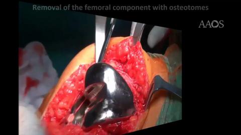
The second step is to remove is the femoral component. The implant-cement or implant-bone interface is identified. The femur is approached from medial to lateral, starting from the anterior flange and moving toward the posterior condyle. Surgeons should maintain parallelism between the saw and the interface to avoid removal of unwanted bone. After the interface is completed with the use of the power saw, the osteotomes can be used to mobilize the femoral component. In patients with a stemmed implant, specific instrumentation may be useful, and an anterior cortical window may be created to remove residual cement.
The last step is to remove the tibial component. The target surface is approached with the use of an oscillating saw from the anterior aspect to the posteromedial corner, with care taken to not damage the surrounding soft tissue. Then, the lateral side can be approached with the use of smaller saws. After the entire tibial platform is completed, stacking, broad osteotomes can be used to detach the component.
Removal of the patellar component should be attempted after thoughtful consideration because of the risk for fracture. The use of osteotomes is discouraged because of the fracture risk. In conclusion, the first step for safe implant removal is to attain good exposure. Implant removal should begin with the polyethylene liner to allow for better exposition for the next steps. In patients with a cemented stem, an anterior cortical window may aid in complete removal. Finally, removal of the patellar component should be carefully planned because of the risk of patellar fracture.